Have you ever found yourself holding a menu at arm’s length to read it clearly? Are you struggling to focus on your phone screen or favorite book? If so, you might be experiencing presbyopia (long-sightedness), a common age-related vision condition. This issue is all too familiar for many people over 40. Presbyopia, or long-sightedness, affects millions and can significantly impact daily life. Understanding what causes presbyopia, recognizing its symptoms, and exploring available treatments can help manage this age-related condition effectively. In this blog, we’ll explore the specifics of presbyopia, providing comprehensive information to help you see life more clearly.
Presbyopia is commonly known as long-sightedness. It is an age-related condition where the eye gradually loses its ability to focus on nearby objects., such as books, mobile screens, or menus.
Unlike other vision problems like myopia (nearsightedness) or hyperopia (farsightedness), presbyopia is not caused by the eyeball’s shape but by the lens’s hardening inside the eye. This change in the lens prevents it from changing shape easily to focus on close objects.
Presbyopia (long-sightedness) eventually affects everyone, even those who have never had vision problems. While it cannot be prevented, it can be managed effectively with the right approach. Understanding presbyopia is the first step towards managing it and maintaining a good quality of life.
Presbyopia (long-sightedness) is a natural result of aging, and its primary cause is the gradual loss of flexibility in the lens of the eye. As we age, the proteins within the lens undergo changes that make the lens harder and less elastic. This loss of elasticity makes it difficult for the lens to change its shape to focus on close objects.
Another contributing factor is the weakening of the ciliary muscles, which help the lens to focus. Over time, these muscles lose their strength, further reducing the eye’s ability to focus on nearby objects.
Another important factor contributing to presbyopia (long-sightedness) is the reduction in pupil size as we age. The pupil is the black circular opening in the center of the iris that allows light to enter the eye. With age, the muscles that control the pupil size become less responsive, causing the pupil to become smaller and less able to dilate effectively in low-light conditions. This reduced ability to adjust the size of the pupil limits the amount of light entering the eye, which can further exacerbate the difficulties in focusing on close objects.
While aging is the main cause of presbyopia (long-sightedness), other factors can influence its onset and severity:
Understanding these causes can help manage presbyopia (long-sightedness) effectively and maintain clear vision as you age.
Common symptoms of presbyopia include:
Recognizing these symptoms and seeking professional eye care can lead to appropriate diagnosis to improve quality of life.
Diagnosing presbyopia (long-sightedness) involves a comprehensive eye examination by an optometrist or ophthalmologist. The examination typically includes:
By carefully assessing the symptoms and conducting these tests, the eye care professional can accurately diagnose presbyopia (long-sightedness) and recommend appropriate treatment options to improve vision.
Presbyopia (long-sightedness) can be managed through various treatment options:
Eyeglasses are a popular and convenient option for correcting presbyopia (long-sightedness).
Contact lenses offer a convenient and effective solution for correcting vision, providing flexibility and comfort for those with presbyopia (long-sightedness).
When it comes to managing presbyopia (long-sightedness) surgically, there are several options available.
Simple eye exercises, like focusing on near and distant objects or rolling the eyes in different directions, can improve flexibility and strength in the eye muscles, enhancing overall vision. Additionally, consuming a diet rich in vitamins and nutrients, particularly those beneficial for eye health like vitamins A, C, and E, as well as omega-3 fatty acids supports eye health.
Each option has its pros and cons, and the most suitable choice depends on factors like age, overall eye health, lifestyle, and personal preferences. It’s crucial to consult with an eye care professional to determine the most appropriate treatment plan.
Effectively managing presbyopia (long-sightedness) in daily life is essential for maintaining clarity and comfort in vision as we age.
Maintain regular eye check-ups to monitor vision changes. Discuss with an eye care professional for personalized recommendations.
Given below is the eye problems list compiled by our expert ophthalmologists
Certain eye conditions and illnesses could have similar symptoms, but others might have distinct indications and symptoms. Here’s a description of the symptoms of eye diseases list given above:
| Eye Conditions | Possible Eye Problems Symptoms |
| Cataracts | cloudy or blurred vision, faded colours, glare, and difficulty seeing at night. |
| Diabetic retinopathy | blurred vision, floaters, fluctuating vision, dark or empty areas in vision, and impaired colour vision. |
| Glaucoma | Often asymptomatic in the early stages; gradual peripheral vision loss, tunnel vision, eye pain, headache, and halos around lights. |
| AMD | blurred or distorted central vision, difficulty reading or recognizing faces, and dark or empty areas in the central vision |
| Retinal detachment | Sudden onset of floaters, flashes of light, and a curtain-like shadow over the visual field. |
| Conjunctivitis | Redness, itchiness, water or discharge, gritty feeling in the eyes, and sensitivity to light. |
| Dry eye syndrome | Stinging or burning sensation, redness, blurred vision, sensitivity to light, and fluctuating vision. |
| Refractive errors | Blurry vision, difficulty seeing at night, eye strain, and headaches. |
| Floaters and flashes | Spots, cobwebs, or specks that float in the field of vision, and flashes of light. |
| Retinitis pigmentosa | Difficulty seeing at night, loss of peripheral vision, and tunnel vision. |
| Keratoconus | Distorted vision, sensitivity to light, and frequent changes in eyeglass prescription. |
| Amblyopia | Reduced vision in one eye, poor depth perception, and misaligned eyes |
| Blepharitis | Red, swollen eyelids, itching, burning, crusty eyelashes, and blurred vision. |
| Strabismus | Misaligned eyes, double vision, and difficulty focusing. |
| Uveitis | Eye pain, redness, blurred vision, light sensitivity, and floaters. |
Even if certain eye problems symptoms could coexist, seeing an eye care specialist for a precise diagnosis and the best course of action is crucial.
In essence, presbyopia (long-sightedness) is a natural part of aging, and with the right approach, it doesn’t have to interfere with your daily life. Regular check-ups and consultations with eye care professionals can help you find the best solution to maintain clear and comfortable vision.
For expert eye care services, consider visiting Skipper Eye-Q International Eye Hospitals. Our experienced professionals are dedicated to providing comprehensive eye care and personalized treatment plans to help you manage presbyopia (long-sightedness) effectively. Book an appointment today and take the first step towards better vision and improved quality of life.


Have you ever noticed tiny spots or threads floating across your vision when looking at a bright sky or a plain wall? Or perhaps you've seen sudden flashes of light,...
Read More
If you're dealing with pain in your right eye along with a headache, it's most often one of a handful of familiar causes a migraine, a tension headache, a sinus...
Read More
Apollo, medically referred to as conjunctivitis occurs from inflammation of the eye's transparent membrane, known as the conjunctiva. This inflammation is triggered by infection or irritation caused by bacterial, viral,...
Read More
Have you ever noticed your parents or family members struggling with diabetes? Perhaps you’ve seen them carefully monitoring their blood sugar levels or diligently taking their medication. But did you know that diabetes can also affect the eyes? This is where diabetic retinopathy comes into play. It’s a common complication of diabetes and can lead to vision loss if left untreated. In this blog, we’ll explore what diabetic retinopathy is, how it affects vision, and what you can do to protect yourself and your loved ones from its consequences. So, let’s dive in and learn more about this important issue together.
Diabetic retinopathy is a condition affecting the eyes, mainly found in people with diabetes. It occurs due to damage to the blood vessels in the retina, which is the light-sensitive tissue at the back of the eye. Over time, high levels of sugar in the blood can weaken and damage the tiny blood vessels that nourish the retina, leading to various complications.
In its early stages, Diabetic Retinopathy may not cause any noticeable symptoms. However, as the condition progresses, it can result in vision problems and even blindness if left untreated. There are two main types of Diabetic Retinopathy.
There are 2 stages of diabetic Retinopathy, as follows:
NPDR is an early stage of diabetic retinopathy. Many humans with diabetes are suffering from it. The small blood vessels leak with NPDR, making the retina swell. If the macula swells, it is called macular oedema. This is the reason why diabetes patients experience vision loss.
Blood vessels will also be closed off by NPDR. This is called macular ischemia. In this condition, blood can’t reach to macula. Sometimes tiny cholesterol particles known as exudates can form in the retina. Early-stage diabetes eye symptoms can affect your vision, too. If you have NPDR, your near vision can be blurry.
PDR is an advanced stage of diabetic retinopathy. It occurs whilst the retina starts off evolving and growing new blood vessels. This is known as neovascularization. These fragile new vessels frequently bleed into the vitreous. If they effectively bleed a little, you may see a few dark floaters. If they bleed a lot, it might block all vision.
These new blood vessels can also form scar tissue. Scar tissue can cause problems with the macula or cause a detached retina. PDR is very critical, and diabetic retinopathy symptoms affect both your vital and peripheral (side) vision.
Awareness of diabetic retinopathy and its potential consequences is essential for individuals with diabetes to take essential steps in managing their condition and protecting their vision.
Here’s a breakdown of the causes of diabetic retinopathy:
Here are some additional factors that can increase your risk of developing diabetic retinopathy:
Understanding these factors can help individuals with diabetes and healthcare providers in implementing preventive measures and early interventions to mitigate the risk of diabetic retinopathy.
Diabetic retinopathy often doesn’t cause noticeable symptoms in its early stages. However, as the condition progresses, symptoms may develop, including:
These symptoms can vary in severity depending on the stage of diabetic retinopathy and other factors.
Anybody with diabetes faces the risks of diabetic retinopathy. However, the risk is higher if the person:
Diabetic patients are at risk of developing diabetic retinopathy. There is a risk of diabetic retinopathy in persons facing type 1, type 2, and gestational diabetes. The risk increases the longer you’ve had diabetes. With time, a huge number of diabetes patients face diabetic retinopathy. By controlling diabetes, you can reduce the risk of diabetic retinopathy.
Pregnant women or women who face gestational diabetes are at high risk of developing diabetic retinopathy. If you are pregnant and a diabetes patient, then you should undergo a dilated eye test as early as possible. Ask our doctor at Skipper Eye-Q International Eye Hospitals if you’ll need more eye tests for the duration of your pregnancy.
Diagnosing diabetic retinopathy involves a comprehensive eye examination by an eye care professional. The exam typically includes dilating the pupils with eye drops to allow for a better view of the retina. The doctor will then use specialized instruments to examine the retina for signs of damage, such as abnormal blood vessels, swelling, or bleeding. They may also perform additional tests, such as optical coherence tomography (OCT) or fluorescein angiography, to assess the extent of retinal damage and guide treatment decisions. Early detection through regular eye exams is key to managing diabetic retinopathy and preventing vision loss.
When it comes to managing diabetic retinopathy, there are several treatment options available to help preserve vision and prevent further damage to the eyes. Here are some common approaches:
Non-invasive treatments for diabetic retinopathy focus on interventions that do not require surgical procedures. These options aim to address retinal damage and prevent further vision loss using techniques such as laser therapy and injections.
Surgical treatments for diabetic retinopathy may be necessary in cases of severe retinal damage or complications such as retinal detachment. These procedures aim to remove blood and scar tissue from the eye to improve vision and prevent further deterioration.
Adjunctive therapies for diabetic retinopathy focus on lifestyle modifications and ongoing management strategies to complement primary treatment approaches and promote overall eye health.
Regular eye exams are essential for monitoring the progression of diabetic retinopathy and detecting any vision changes. Ophthalmologists can assess the effectiveness of treatment and make adjustments as necessary to preserve vision and overall eye health.
Collaborative care involves working closely with a team of healthcare professionals, including ophthalmologists, endocrinologists, and primary care physicians, to ensure comprehensive management of diabetic retinopathy. This collaborative approach helps address various aspects of the condition and optimize treatment outcomes.
Preventing diabetic retinopathy involves managing diabetes effectively and adopting healthy lifestyle habits. Here are key preventive measures:
By following these preventive measures, you can significantly protect your vision.
Living with diabetic retinopathy requires proactive management and regular medical care. Regular eye exams are essential for monitoring the condition and detecting any changes early. Adhering to prescribed treatment plans and seeking prompt medical attention for any vision changes or complications is crucial. With proper care and attention, individuals can maintain their vision and quality of life despite diabetic retinopathy.
Given below is the eye problems list compiled by our expert ophthalmologists
Certain eye conditions and illnesses could have similar symptoms, but others might have distinct indications and symptoms. Here’s a description of the symptoms of eye diseases list given above:
| Eye Conditions | Possible Eye Problems Symptoms |
| Cataracts | cloudy or blurred vision, faded colours, glare, and difficulty seeing at night. |
| Diabetic retinopathy | blurred vision, floaters, fluctuating vision, dark or empty areas in vision, and impaired colour vision. |
| Glaucoma | Often asymptomatic in the early stages; gradual peripheral vision loss, tunnel vision, eye pain, headache, and halos around lights. |
| AMD | blurred or distorted central vision, difficulty reading or recognizing faces, and dark or empty areas in the central vision |
| Retinal detachment | Sudden onset of floaters, flashes of light, and a curtain-like shadow over the visual field. |
| Conjunctivitis | Redness, itchiness, water or discharge, gritty feeling in the eyes, and sensitivity to light. |
| Dry eye syndrome | Stinging or burning sensation, redness, blurred vision, sensitivity to light, and fluctuating vision. |
| Refractive errors | Blurry vision, difficulty seeing at night, eye strain, and headaches. |
| Floaters and flashes | Spots, cobwebs, or specks that float in the field of vision, and flashes of light. |
| Retinitis pigmentosa | Difficulty seeing at night, loss of peripheral vision, and tunnel vision. |
| Keratoconus | Distorted vision, sensitivity to light, and frequent changes in eyeglass prescription. |
| Amblyopia | Reduced vision in one eye, poor depth perception, and misaligned eyes |
| Blepharitis | Red, swollen eyelids, itching, burning, crusty eyelashes, and blurred vision. |
| Strabismus | Misaligned eyes, double vision, and difficulty focusing. |
| Uveitis | Eye pain, redness, blurred vision, light sensitivity, and floaters. |
Even if certain eye problems symptoms could coexist, seeing an eye care specialist for a precise diagnosis and the best course of action is crucial.
It’s crucial to raise awareness about diabetic retinopathy to safeguard vision. Prioritize eye health and seek early treatment if you have diabetes. For those seeking specialized care and treatment for diabetic retinopathy, reach out to Skipper Eye-Q International Eye Hospitals. With their expertise and dedication to eye care, they can provide the necessary support and guidance to manage diabetic retinopathy effectively. Don’t hesitate to take the first step towards preserving your vision and ensuring a brighter future for your eyes.
No, Diabetic retinopathy can not be reversed. But its growth is slowed or prevented by proper management. Early-stage diabetic retinopathy may improve with strict blood sugar, LDL cholesterol management and blood pressure. But if you face major damage, then you undergo treatments like laser therapy, anti-VEGF injections, or surgery that can be required to prevent further vision loss.
Diabetic retinopathy is detected through a complete eye test, which incorporates dilated eye exams, fundus photograph, optical coherence tomography (OCT) scans, and fluorescein angiography. During the dilated eye tests, an ophthalmologist examines the retina for atypical blood vessels, bleeding, or swelling. Regular screenings are essential for individuals with diabetes, as early detection can save them from extreme situations.
In the early stages, diabetic retinopathy might not cause noticeable diabetic retinopathy symptoms. However, the main signs and symptoms frequently include blurred vision, issues seeing at night, and the appearance of floaters or dark spots in the vision. As the condition progresses, vision might also become increasingly distorted, and untreated cases can cause blindness. Regular eye checkups help in figuring out and handling the condition before intense damage occurs.
No. It will not heal on its own. Making life adjustments in the early stages may reverse it; however, later stages require treatment. You want to see our health practitioner at Skipper Eye-Q International Eye Hospitals for routine exams. They can test for diabetes eye symptoms of the disease and discover the best treatment option.
Once your medical doctor has identified the disease and the way it is, they will create a tailor-made treatment plan. Diabetic retinopathy treatment consists of Laser approaches, injection of drugs which could address both leakage and bleeding in the retina (known as anti-angiogenic injections), and a vitrectomy surgery to remove the clear gel (vitreous) that fills the centre a part of your eye.
Have you ever noticed tiny spots or threads floating across your vision when looking at a bright sky or a plain wall? Or perhaps you've seen sudden flashes of light,...
Read More
If you're dealing with pain in your right eye along with a headache, it's most often one of a handful of familiar causes a migraine, a tension headache, a sinus...
Read More
Apollo, medically referred to as conjunctivitis occurs from inflammation of the eye's transparent membrane, known as the conjunctiva. This inflammation is triggered by infection or irritation caused by bacterial, viral,...
Read More
The majority of people suffer from cataract, which manifests as an opaque or clouded lens in either one or both eyes. Fortunately, there is a less invasive eye cataract operation that can reverse cataracts and the resulting sight loss.
The lens is translucent and flexible when you are born, directing lights onto the retina located at the rear of the eye. A distinct view is produced by this focus. However, as we age, proteins in our lenses clump together and impair our vision, making it harder to discern details. An eye cataract operation is the solution for that.
It is natural to have post-procedure doubts and say there are so many things I wish I knew before cataract surgery. Despite its safety and high success rate, anxiety in patients undergoing cataract surgery is very common. Anxiety associated with cataract surgery is one of the main reasons candidates for cataract surgery delay surgery. At Skipper Eye-Q Super Specialty Eye Hospital, we encourage our patients not to let the fear and anxiety of surgery increase worry.
Must Read – Cataracts: Causes, Symptoms, Treatment
This post will go over twenty facts that you should know before having cataract surgery to help you feel more at ease and less nervous.
Before we get to the surgery tips, it helps to know the cataract symptoms that tell you it may be time to see an eye doctor. A cataract grows slowly, so many people do not notice it at first. The clouding builds up over months or years.
The most common signs of a cataract are:
If two or three of these sound familiar, book a comprehensive eye examination. You can also read our full guide on cataracts: causes, symptoms and treatment to learn more.
Among the safest and most common surgical procedures carried out globally is cataract surgery. Every year, millions of patients get this operation to help them see again.
To evaluate the best plan of action and identify the extent of your cataracts, your eye specialist will do a thorough examination of your eyes before the operation. In addition, they will give you advice on how to be ready for the operation, such as what drugs to avoid taking and when to cut back on food and liquids.
There are several forms of cataract surgery, such as laser-assisted surgeries and conventional phacoemulsification. Based on your specific requirements and the state of your eyes, your doctor will advise you on the best eye cataract operation.
Local anaesthesia is usually used during cataract surgery, so although you will be awake, your eye is under anaesthesia to minimise any discomfort.
Most patients can go back home the same day after the procedure, which typically takes 15 to 30 minutes for each eye.
Post cataract surgery, most patients report a noticeable improvement in their eyesight. To attain the best possible visual acuity, you might need to wear eyeglasses or contact lenses while waiting for your eyes to fully adapt.
You shouldn’t experience any pain throughout the procedure; instead, you should just feel a little pressure or motion in your eye. Tell your surgeon right away if you feel any discomfort.
Although the procedure is brief, recuperating from cataract surgery might take a few weeks. In the days after surgery, you can have light sensitivity, hazy vision, and minor pain.
Infection and haemorrhage are two concerns associated with cataract surgery, just like with any surgical operation. Serious cataract operation risks are uncommon, though, and surgery’s advantages frequently exceed its drawbacks.
Post cataract surgery, you might need to make some adjustments to your way of life, such as avoiding physically demanding tasks and wearing sunglasses outside to shield your eyes from UV rays. You must adhere to your doctor’s advice to get the greatest possible result.
Must Read : Red Eye After Cataract Surgery? Foods to Avoid
Toric, multifocal, and mono-focal intraocular lenses (IOLs) are among the several types of cataract lenses that can be used during surgery. Talk with your surgeon to find the best solution for your requirements and lifestyle since each kind has its benefits and considerations.
Although cataract surgery is usually safe, there are always certain associated cataract operation risks. These might include inflammation, detachment of the retina, and oedema. Before the treatment, your surgeon will go over these risks with you and take precautions to reduce them.
You can have minor pain, glare, halos around lights, and impaired vision as a transient cataract operation after effects following surgery. As your eyes heal, these sensations usually get better in a couple of days to weeks.
Your eyes may need some time to properly adjust to the cataract surgery, even though many people report considerable improvements in their vision quickly after the procedure. Over a few weeks or months, while your eyes recover and adjust to the new implanted lens, your vision can keep progressively improving.
Until your eyesight has stabilised and your surgeon gives the all-clear to resume driving, you must refrain from driving right after cataract surgery.
To track your healing post cataract surgery and guarantee the best possible visual results, you will need to schedule routine follow-up visits with your surgeon. To encourage recovery and avoid problems, make sure you adhere to all post-operative care recommendations given by your surgeon.
To lower the cataract operation risks of elevated intraocular pressure or dislodging the intraocular lens, it is imperative to avoid vigorous activity, heavy lifting, and bending over in the days after cataract surgery.
Cataract surgery has a very high patient satisfaction rate and is a very successful technique. Post cataract surgery, the majority of patients have greater vision and a longer-lasting quality of life.
Although health insurance frequently covers cataract surgery, it’s important to check with your physician to find out what your coverage entails and how much you’ll pay out of cash.
Before having cataract surgery, it’s common to have anxiety or nervousness. Don’t be afraid to ask for emotional assistance and comfort at this time from relatives, close friends, or support groups.
Must Read – How Long Should I Wait To Wash My Hair After Cataract Surgery?
Before your cataract surgery, your doctor will recommend a complete eye test to assess the severity of your cataracts and decide the precise treatment plan depending on the cataract stage. In the early stages, cataracts are treated with glasses. Once it interferes with daily activities, you need to undergo surgery.
Besides selecting what type of cataract surgery you want to have, you should choose an intraocular lens (IOL) implant to replace the natural lens so that it can be removed during the process. Your vision final results will range extensively, relying on the IOL you selected, so it’s recommended to consult your doctor to understand things I wish I knew before cataract surgery.
Cataract surgery is a short, painless outpatient system that commonly takes 15–20 minutes per eye. Here’s a short analysis of things I wish I knew before cataract surgery
Here are the steps you should know to prepare for a cataract surgery
Before your surgery, you’ll have a comprehensive session at our Skipper Eye-Q International Eye Hospitals. During this appointment, our ophthalmologists will conduct an intensive eye examination and talk about your clinical records to decide the best approach to your cataract surgery.
To ensure the success of your Cataract surgery, follow the given preoperative instructions.
Must Read – 15 Tips on How to Train Your Eyes After Cataract Surgery
Cataract surgery is one of the safest operations in the world, with a success rate of around 98%. But honest patients will tell you there are a few things they wish someone had explained more clearly beforehand. Knowing the disadvantages and the common problems ahead of time helps you set the right expectations — and that is the single biggest reason people end up happy with their results.
Most problems are mild and temporary. The usual ones are blurry vision for a few days, light sensitivity, mild grittiness, watery eyes, and the glare or halos mentioned above. Serious complications — like infection or retinal detachment — are rare, happening in well under 1 in 100 cases. Following your eye drop schedule and avoiding rubbing your eye keeps the risk low.
Genuine regret is uncommon, but when it happens it usually comes down to expectations, not the surgery itself. The biggest reasons people feel let down are:
The fix is simple: have an honest conversation about your daily life — driving, reading, screens, hobbies — before you pick a lens. A good surgeon will match the lens to your routine. Book a consultation and ask every question on your mind. There is no such thing as a silly question about your own eyes.
Although most of the patients experience smooth recovery, this surgery has some warming symptoms. Few of them are listed below:-
Most patients experience smooth recovery, however be alert for potential warning symptoms.
Normal in the first week, but vision needs to be enhanced step by step.
Small shadows or specks in vision are common however, they must fade regularly.
Seek on-the-spot medical help if you experience problems like contamination or retinal detachment.
Could sign an infection—contact with your eye hospital if this takes place.
Hope after reading these knowledgeable facts by our experts you’re not still wondering about things I wish I knew before cataract surgery. At Skipper Eye-Q Super Specialty Eye Hospital, we strive to improve the quality of life for our patients by using cutting-edge eye cataract operation and vision correction techniques. People of all ages can benefit from modern vision correction procedures performed by our team of skilled ophthalmologists. You may quickly go to clear vision by contacting our office to schedule your thorough eye exam and consultation.
Cataract surgery is a treatment procedure in which your eye lenses are removed and replaced with artificial lenses. A cataract causes the lens to become cloudy while it’s usually clear. Cataracts can affect vision.
Cataract surgery is accomplished through our eye health practitioner at Skipper Eye-Q International Eye Hospitals. It’s accomplished on an outpatient basis, so you don’t need to stay in the hospital after the surgery. Cataract surgery is very common and is commonly a safe surgery.
The three main types of cataracts are nuclear cataracts (which form in the centre of the lens and are the most common with age), cortical cataracts (which form as wedge-shaped streaks on the outer edge of the lens), and posterior subcapsular cataracts (which form at the back of the lens and often affect reading and night vision faster). An eye exam will show which type you have.
Cataract surgery has a success rate of around 98%, making it one of the safest and most successful operations performed anywhere in the world. The vast majority of patients see a clear improvement in their vision. Serious complications happen in less than 1 in 100 cases, especially when the surgery is done by an experienced eye surgeon.
The main disadvantages are that you may still need glasses for some tasks with a standard lens, premium lenses cost more, and some people notice glare or halos at night for a few weeks. Dry eyes are common early on, and a thin film can cloud behind the lens months later (easily cleared with a quick laser). Serious risks are rare.
Most surgeons wait about 1 to 4 weeks between operating on each eye, with 1 to 2 weeks being common. This gives the first eye time to settle and lets your surgeon confirm the lens power is right before doing the second eye. Your surgeon will set the exact gap based on how your first eye heals.
The most feared complication is endophthalmitis, a serious infection inside the eye. It is very rare, affecting far fewer than 1 in 1,000 patients, and is the reason antibiotic eye drops and strict sterile steps are used. Other rare complications include retinal detachment and swelling at the back of the eye. Prompt treatment usually protects vision.
Toric lenses correct astigmatism, but they must sit at an exact angle. If a toric lens rotates out of position, vision can blur and a small procedure may be needed to reposition it. Some patients also notice mild glare or still need a light glasses prescription. Choosing an experienced surgeon lowers the chance of these problems.
Most people see more clearly within 24 to 48 hours, but some blurriness is normal for the first few days to a week. Vision keeps sharpening over 4 to 8 weeks as the eye fully heals and settles around the new lens. If blurriness suddenly gets worse instead of better, contact your eye doctor.
Before cataract surgery, you should avoid eating or drinking anything for at least 6 to 8 hours, as instructed by your eye specialist. Avoid wearing makeup, creams, or lotions around your eyes on the day of the procedure. Additionally, consult your ophthalmologist about stopping any medications, especially blood thinners, as they might increase the risk of bleeding during surgery.
The most common complaint after cataract surgery is mild discomfort, including a gritty or scratchy feeling in the eye. Some patients may also notice blurry vision for a few days as the eye adjusts to the new lens. Dry eyes, light sensitivity, and halos around lights at night are also temporary complaints that typically resolve within a few weeks. Using prescribed eye drops and following post-surgery instructions recommended by your ophthalmologists can help minimize these symptoms.
Resting for 1 to 2 days after cataract surgery is essential. While most patients can resume light activities within 24 to 48 hours, it is recommended to avoid strenuous activities, heavy lifting, or swimming for at least a week. Following your ophthalmologist’s advice ensures a smooth recovery and reduces the risk of complications.
Though cataract surgery is highly successful, the biggest risks include minute infections, inflammation, or retinal detachment. These complications are rare, occurring in less than 1% of cases. At Skipper Eye-Q, we prioritize patient safety by using state-of-the-art equipment and strict hygiene protocols to minimize risks and ensure successful outcomes.
Avoid eating spicy, oily, or salty foods immediately after cataract surgery, as they can increase inflammation or discomfort. Foods high in sugar or caffeine should also be limited to reduce the risk of irritation or dehydration. Instead, focus on a balanced diet rich in fruits, vegetables, and foods high in omega-3 fatty acids to promote faster healing and maintain overall eye health.
It is strictly recommended that you use the eye drops before your cataract surgery. If you don’t use the eye drops, then you definitely don’t get the exact final result after surgery. Not using eye drops can increase the recovery time and postpone the healing process. It may also increase the risk of infection or irritation.
Cyclopentolate and Phenylephrine are recommended for use before cataract surgery. These eye drops are medicinal drugs that help you recover and keep your eyes healthy. Eye drops can blur the vision for a while, approximately.
It is recommended that you take medicine with the guidance of your health practitioner at Skipper Eye-Q International Eye Hospitals, as it’s possible that these medicines can affect cataract surgery. Some antibiotic eye drops are beneficial to apply 1-2 days before the cataract surgery.
It is recommended to stay aware of scared of cataract surgery, stop taking a few aspirin and anti-clotting drugs as there might be slicing of eye lenses so small bleeding can arise. Thus, you should follow the guidelines prescribed by your physician.
Cataract surgery recovery takes about 4 to 8 weeks. While you can see through the operated eye in about 24 hours, it takes 1 to 2 months for the eye to fully heal. There are some restrictions due to light sensitivity and pressure on the operated area. However, you can view the world normally through the operated eye while wearing protective eyewear. You may experience blurry or hazy vision with light sensitivity and mild irritation in the beginning.
Any strenuous activity is strictly restricted after a cataract surgery. You should avoid exercising and any work that requires you to bend down or lift heavy objects. It is generally allowed to read, watch television, or use your phone. However, make sure not to put too much strain on your eyes. Also, avoid randomly touching the operated eye or rubbing it.
You should not rub your eyes until your cataract surgery recovery time is over. Putting pressure on the surgical wound can disrupt the healing process and might cause unexpected complications. The cataract surgery recovery takes about 1 to 2 months in most cases. During this time, it is crucial to treat the operated eye with special care.
It is recommended to avoid washing your hair for 24 to 48 hours after a cataract surgery. Later on, wash your hair with your head tilted back. Take care that the water or shampoo does not go into your eyes. Use a mild shampoo and gently massage your scalp. Rinse thoroughly and gently, and dry your hair while still keeping your head tilted back.
Have you ever noticed tiny spots or threads floating across your vision when looking at a bright sky or a plain wall? Or perhaps you've seen sudden flashes of light,...
Read More
If you're dealing with pain in your right eye along with a headache, it's most often one of a handful of familiar causes a migraine, a tension headache, a sinus...
Read More
Apollo, medically referred to as conjunctivitis occurs from inflammation of the eye's transparent membrane, known as the conjunctiva. This inflammation is triggered by infection or irritation caused by bacterial, viral,...
Read More
Have you had cataract removal surgery done recently, or are you thinking about having one? At Skipper Eye-Q Super Specialty Eye Hospital, our eye doctors offer excellent advice to hasten the healing of your eyes. Depending on your circumstances and how well you follow surgical instructions, recovery might take up to two months. Getting your eyes used to doing regular chores is one of the simplest strategies for dealing with your vision imbalance.
You may be confident that cataract surgery is both safe and very successful, particularly if you visit a reputable eye hospital like Skipper Eye-Q Super Speciality Eye Hospital. Still, how long does it take to heal from cataract surgery depends on the postoperative work.
Training your eyes after cataract surgery might help you heal faster. Our team of cataract surgeons has compiled important material, which includes activities to help your eyes get used to the replacement lens implants.
A cataract treatment is a correction of vision using laser surgery that involves the removal of your native eye lens and replacement with an artificial one. Your eye surgeon will assist you in determining which of the many lenses available will best meet your visual objectives.
You may leave the outpatient procedure a few hours later, with each eye taking fifteen to twenty minutes to operate on. After the surgery, our team will keep an eye on your recovery, respond to any concerns you might have, and give you thorough instructions on how to take care of yourself thereafter.
Adhering to the recommended postoperative protocols will determine, in part, excellent outcomes and how long does it take to heal from cataract surgery. To achieve the greatest outcomes and a pleasing visual effect, do the tasks listed below. You can return to your regular activities more quickly by using your surroundings as a training ground (without glasses or contacts!).
These are the 15 tips for training your eyes after cataract surgery:
Following a cataract treatment surgery, you may enhance your eyesight and promote a speedy recovery by adhering to your post-operative care regimen and these guidelines. During your recuperation, don’t forget to contact your eye doctor with any queries or if you encounter any strange symptoms.
It will be simpler to do the exercise by yourself now that you are aware of a few techniques for training your eyes after cataract surgery. Recall that engaging in these activities can aid in hastening your recuperation. Having the operation is already the culmination of your hard work. To get the greatest outcomes possible, it’s crucial to do these last tasks.
Stop right away if any of the above actions make you uncomfortable or if you have any further queries. If you feel pain or discomfort while performing these exercises, speak with your eye doctor before continuing. Depending on your medical background and present state of vision, your eye doctor can recommend a different set of exercises for you to perform after your cataract removal.
You can count on Skipper Eye-Q Super Speciality Eye Hospital to support you at every step of the process, from preoperative consultations to actual cataract treatment and beyond. Our cataract surgery experts provide the greatest care possible in terms of fostering a caring, supporting atmosphere and attending to your overall health and well-being.
When it comes to returning to your regular activities, heed the advice of your physician. A prescription from your doctor for eye drops and anti-infection drugs will be given to you. Additionally, our staff will educate you on things to avoid while recovering. We can promptly assess any potential surgical problems at every follow-up visit. If you still require spectacles or contact lenses, don’t forget to take benefit of our other offerings.
Our goal at Skipper Eye-Q Super Speciality Eye Hospital is to work with you to have the greatest eyesight possible following refractive cataract surgery. Together, we can choose the best care and treatments to help you see better.
Get in touch with us right now to arrange a consultation or to find out more about training eyes after cataract surgery. Our compassionate eye specialists will support you both during and after your cataract-removal surgery.
Have you ever noticed tiny spots or threads floating across your vision when looking at a bright sky or a plain wall? Or perhaps you've seen sudden flashes of light,...
Read More
If you're dealing with pain in your right eye along with a headache, it's most often one of a handful of familiar causes a migraine, a tension headache, a sinus...
Read More
Apollo, medically referred to as conjunctivitis occurs from inflammation of the eye's transparent membrane, known as the conjunctiva. This inflammation is triggered by infection or irritation caused by bacterial, viral,...
Read More
The terms “itchy eyes” or “dry eyes” describe a condition in which there is insufficient lubrication or moisture in the eyes, causing pain, itching, and irritation. Environmental factors, extended use of screens, ageing, and specific medical disorders are common reasons for itchy watery eyes. To provide the best possible eye health and comfort, Skipper Eye-Q International Eye Hospitals offer comprehensive care and cutting-edge treatments for a range of eye diseases, including dry eyes and itchy eyes.
Typical signs and symptoms include:
There are a variety of potential itchy eyes causes including :
For dry eyes or itchy eyes to be effectively managed and relieved, the underlying itchy eyes causes must be found.
Must Read – Dry Eye Syndrome: Causes, Symptoms, & Treatment
Home remedies for dry eyes provide all-natural ways to reduce irritation and enhance general eye health.
Must Read – Top 15 Common Eye Disorders and Diseases
If you don’t have eye drops on hand, a few of the remedies above work quickly on their own:
These steps won’t replace treatment for an underlying condition, but they’re the quickest drop-free way to settle occasional itchiness.
Itchy eyes and watery eyes often happen together, but they don’t always share the same cause, so it helps to tell them apart:
Rule of thumb: allergy-driven watering responds fastest to cold compresses and allergen avoidance; dryness-driven watering responds better to lubrication and rest breaks.
Several remedies already used for generations in Nigeria households line up well with the natural options above:
A note on safety: these are complementary, not curative — they ease mild irritation but won’t treat an infection, allergy, or dry eye condition on their own. Never put any of these substances (including rose water or milk) directly inside the eye; use them only on closed eyelids, and discontinue if irritation
To relieve symptoms and encourage healing, a variety of therapies may be recommended in cases of severe itchy watery eyes:
In cases of extremely dry and itchy eyes, these therapies are intended to relieve the immediate symptoms and improve ocular health. The best course of therapy should be decided upon in consultation with the knowledgeable ophthalmologists at Skipper Eye-Q International Eye Hospitals, taking into account each patient’s unique needs and the severity of their symptoms.
Must Read – Home Remedies for Itchy Eyes
Here are some more helpful techniques that might improve comfort and encourage relief:
1. Protection Against Environmental Irritants: Dryness and itching can be considerably exacerbated by environmental variables.
Wear wrap-around shades when you’re outside to shield your eyes from dust, wind, and pollen, all of which can cause irritation and increased tear production. Steer clear of smoky or perfumed areas, such as those with burning incense or scented sprays, as they can exacerbate eye dryness and itching. These barriers reduce discomfort and preserve moisture.
2. Take Regular Screen Time Breaks: Many individuals spend hours staring at screens without blinking sufficiently, which unintentionally exacerbates dryness. Screens slow down blinking, which causes irritation and tear evaporation. Simple methods consist of:
a) The 20-20-20 rule states that you should gaze at an item at least 20 feet away for 20 seconds every 20 minutes.
b) Blink exercises: To evenly distribute your tears, gently close and open your eyes multiple times while using a screen.
c) To avoid the need to open your eyes wide and avoid tear film tension, place screens a little below eye level.
3. Keep the optimum Indoor Humidity: Dry indoor air, particularly from heaters or air conditioners, can quickly erode your natural tear film, making dry eyes worse. By adding moisture to the air, a humidifier in your bedroom or office reduces tear evaporation and eases irritation. This approach is particularly helpful during the dry or winter months.
4. Eating and Drinking Right to Promote Eye Health: Tear production is more influenced by nutrition and hydration than most people realize. Increase Water intake as tear volume is supported by enough hydration.
Omega-3 fatty acids: Found in walnuts, flaxseeds, chia seeds, and fatty fish (such as salmon and sardines), these fats aid to enhance the oil layer of tears by lowering itching and evaporation.
5. Gently and frequently clean your eyelids: A healthy tear film is maintained by the meibomian glands, which are oil glands located at the edges of the eyelids. Dryness and discomfort may intensify if these glands are plugged. A mild eyelid hygiene regimen can help these glands:
6. Break from wearing Contact lenses: Dryness and itching can be greatly exacerbated by contact lenses, particularly in eyes that are sensitive or allergic. Until your symptoms improve, take breaks from contacts and switch to glasses.
To avoid irritation or contamination, do not apply eye drops right before putting in lenses. If your eye care professional advises it, choose scleral lenses, which are bigger lenses that vault over the cornea and retain moisture.
7. When Basic Home Care Is Insufficient: Dry eye syndrome or another ailment, like meibomian gland malfunction or allergic conjunctivitis, may be the cause of persistent dryness and itching despite screen breaks, hydration, and good hygiene. Because chronic dry eye may require prescription medications or specialist therapy, persistent symptoms should be evaluated by a professional.
These extra helpful added home care tips, which center on daily routines, surroundings, diet, and eye care practices, provide more assured and trusted resources than just compresses and water. They aid in addressing the root causes of dry and itchy eyes as well as their symptoms.
Dry and itchy watery eyes can be relieved with a few simple steps in addition to prescription treatments. To lessen eye strain during the day, these include taking frequent breaks from screens, avoiding smoking and windy conditions, and using a humidifier to bring moisture to the air. Using protective eyewear when participating in activities that might cause red eyes and donning wrap-around shades to shield the eyes from strong sunlight can also help reduce symptoms. Including these easy techniques in everyday activities can improve general eye health and comfort.
With Skipper Eye-Q International Eye Hospitals, find lasting relief from dry or itchy eyes. Our skilled ophthalmologists provide individualised care based on your requirements. Avoid letting ocular pain lower your standard of living. Make an appointment for a consultation right now to learn practical strategies for dry eyes and long-term eye health and comfort.
Home remedies are usually enough for occasional itchiness, but book an eye check if you notice any of the following:
Persistent or worsening symptoms can point to conditions like blepharitis, allergic or infective conjunctivitis, or meibomian gland dysfunction all of which respond much better to early treatment. A comprehensive eye examination can pinpoint the cause. Book a consultation with Skipper Eye-Q’s ophthalmologists if any of the above apply to you.
To quickly alleviate itchy eyes, try using over-the-counter artificial tears or lubricating eye drops. They can help wash away irritants and keep your eyes moist. Applying a cold compress can also provide immediate relief by reducing inflammation and soothing the itch. If your itchy eyes are due to allergies, taking an antihistamine can help control the symptoms.
Saltwater, or saline solution, can help cleanse the eyes by flushing out irritants and allergens. It’s gentle on the eyes and can provide temporary relief from itching. You can purchase sterile saline solution from a pharmacy or prepare a mild saline solution at home by mixing one teaspoon of salt in a liter of distilled or boiled (then cooled) water. However, avoid using too much salt or unclean water, as this can cause further irritation.
Rubbing your eyes can worsen the irritation and even damage the delicate tissues. To stop the habit, try these tips:
Yes, ice can help relieve itchy eyes. The cold temperature reduces inflammation and numbs the area, providing temporary relief from itching. You can wrap some ice cubes in a clean cloth and apply them to your closed eyelids for a few minutes. This can help reduce swelling and soothe the discomfort.
It’s not advisable to apply ice directly to your eyes, as it can cause frostbite or damage the delicate skin around your eyes. Instead, always wrap the ice cubes in a clean, soft cloth before applying them to your closed eyelids. This method will provide the benefits of the cold temperature without the risk of injury.
A cold compress is the fastest drop-free option press a chilled, damp cloth over closed eyes for 5–10 minutes. Rinsing with cool water, avoiding rubbing, and resting your eyes away from screens also help calm itching without any product.
Identify whether the trigger is allergy-related (pollen, dust, pet dander) or dryness-related (screens, dry air). For allergies, a cold compress plus avoiding the allergen works fastest. For dryness, blinking exercises and lubricating drops are more effective. See a doctor if both symptoms persist beyond a few days.
See an eye doctor if itching lasts more than 2–3 days, is accompanied by discharge, pain, blurred vision, or eyelid swelling, or started after an injury or while wearing contact lenses these can signal an infection or condition that needs treatment beyond home care.
Have you ever noticed tiny spots or threads floating across your vision when looking at a bright sky or a plain wall? Or perhaps you've seen sudden flashes of light,...
Read More
If you're dealing with pain in your right eye along with a headache, it's most often one of a handful of familiar causes a migraine, a tension headache, a sinus...
Read More
Apollo, medically referred to as conjunctivitis occurs from inflammation of the eye's transparent membrane, known as the conjunctiva. This inflammation is triggered by infection or irritation caused by bacterial, viral,...
Read More
Eye diseases include a wide range of ailments that impact different areas of the eye, such as the retina and cornea. If treatment for these conditions is not received, vision problems or even blindness may result. Comprehending the various categories of ocular disorders and eye diseases that cause blindness is essential for timely identification, efficient remediation, and successful handling.
Every illness, from common ones like glaucoma and cataracts to uncommon ones like ocular melanoma, needs specialized treatment based on its unique features and course. All of our patient’s vision is preserved and improved by Skipper Eye-Q International Eye Hospitals, from precise diagnosis to cutting-edge treatment methodologies. Our skilled ophthalmologists are dedicated to providing the best possible treatment for eye issues to help you keep your best possible vision.
Given below is the eye problems list compiled by our expert ophthalmologists
Must Read – What is the Most Common Eye Disease in Africa?
Certain eye conditions and illnesses could have similar symptoms, but others might have distinct indications and symptoms. Here’s a description of the symptoms of eye diseases list given above:
| Eye Conditions | Possible Eye Problems Symptoms |
| Cataracts | cloudy or blurred vision, faded colours, glare, and difficulty seeing at night. |
| Diabetic retinopathy | blurred vision, floaters, fluctuating vision, dark or empty areas in vision, and impaired colour vision. |
| Glaucoma | Often asymptomatic in the early stages; gradual peripheral vision loss, tunnel vision, eye pain, headache, and halos around lights. |
| AMD | blurred or distorted central vision, difficulty reading or recognizing faces, and dark or empty areas in the central vision |
| Retinal detachment | Sudden onset of floaters, flashes of light, and a curtain-like shadow over the visual field. |
| Conjunctivitis | Redness, itchiness, water or discharge, gritty feeling in the eyes, and sensitivity to light. |
| Dry eye syndrome | Stinging or burning sensation, redness, blurred vision, sensitivity to light, and fluctuating vision. |
| Refractive errors | Blurry vision, difficulty seeing at night, eye strain, and headaches. |
| Floaters and flashes | Spots, cobwebs, or specks that float in the field of vision, and flashes of light. |
| Retinitis pigmentosa | Difficulty seeing at night, loss of peripheral vision, and tunnel vision. |
| Keratoconus | Distorted vision, sensitivity to light, and frequent changes in eyeglass prescription. |
| Amblyopia | Reduced vision in one eye, poor depth perception, and misaligned eyes |
| Blepharitis | Red, swollen eyelids, itching, burning, crusty eyelashes, and blurred vision. |
| Strabismus | Misaligned eyes, double vision, and difficulty focusing. |
| Uveitis | Eye pain, redness, blurred vision, light sensitivity, and floaters. |
Even if certain eye problems symptoms could coexist, seeing an eye care specialist for a precise diagnosis and the best course of action is crucial.
Depending on the individual state, there might be wide variations in the reasons for eye problems and diseases. Certain illnesses could have similar root causes, but there could be differences as well:
Cataracts: Usually brought on by aging-related changes in the eye’s lens, although they can also result from trauma, certain drugs, or underlying illnesses like diabetes.
Diabetic retinopathy: A condition brought on by diabetes that damages the blood vessels in the retina.
Glaucoma: Usually brought on by elevated intraocular pressure, however, other causes such as inadequate blood supply to the optic nerve can also cause this condition.
AMD: The precise origin is unknown, however age, heredity, food, and smoking are risk factors.
Retinal detachment: Caused by the retina’s separation from the tissue underneath, retinal detachment is frequently brought on by age, trauma, or underlying eye disorders.
Conjunctivitis: Allergies, irritants, and infections caused by bacteria or viruses can all result in conjunctivitis.
Dry eye syndrome: Age, hormonal fluctuations, drugs, environmental variables, or underlying medical issues are some of the causes.
Refractive errors: Due to variations in the ocular structure, these lead to fuzzy vision.
Floaters and flashers: Usually brought on by aging-related alterations to the vitreous humor of the eye, can also be brought on by specific medical diseases or ocular trauma.
Retinitis pigmentosa: Typically brought on by changes in the retina’s DNA.
Keratoconus: Environmental and genetic factors may have a role.
Amblyopia: Usually brought on by aberrant vision development during early life.
Blepharitis: Eyelid gland dysfunction or bacteria are common causes.
Strabismus: Usually brought on by an imbalance in the muscles controlling the eyes.
Uveitis: Infections, autoimmune diseases, and inflammatory conditions.
Every illness in the eye diseases list has its distinct collection of contributing elements, even if some causes may be similar. An expert in eye care must be consulted for a thorough assessment and suitable treatment.
Must Read – 10 Tips for Avoiding Eye Infections
Treatment for eye diseases varies greatly according to the particular ailment, how severe it is, and personal characteristics including general well-being and medical background.
Cataracts: Surgery is typically required to remove the clouded lens and substitute it with a prosthetic intraocular lens.
Diabetic retinopathy: To avoid vision loss, it is managed using control of blood sugar, laser treatment, injections, or surgery.
Glaucoma: To lower intraocular pressure and stop more visual nerve damage, treatment options for the condition may include surgery, eye drops, oral drugs, laser therapy, or other procedures.
AMD: To stop the disease’s development and maintain vision, treatments for the condition include photodynamic therapy, laser therapy, and anti-VEGF injections.
Retinal detachment: To reconnect the retina and avoid irreversible vision loss, emergency surgery is frequently necessary.
Conjunctivitis: Treatment options vary depending on the root cause and may involve the use of antibiotics, antiviral drugs, or antihistamines.
Dry eye syndrome: Treatment options include prescription eye drops, artificial tears, lifestyle changes, and tear outflow blockage surgeries.
Refractive errors: Contact lenses, glasses, or refractive surgery like LASIK can be used as treatments.
Floaters and flashes: Treatment is often not essential unless they significantly impair vision or signal a retinal tear or detachment, that could require immediate medical intervention.
Retinitis pigmentosa: Although there isn’t a cure at this time, there are various treatment options, such as vitamin pills, low vision aids, and gene therapy.
Keratoconus: Treatment options include corneal transplant surgery, collagen cross-linking, and special contact lenses.
Amblyopia: Using vision treatment, covering the stronger eye to reinforce the weaker one, or donning spectacles are common forms of treatment.
Blepharitis: Antibiotics, anti-inflammatory drugs, warm compresses, and good eyelid cleanliness may all be used as treatments.
Strabismus: Treatment options involve vision therapy, spectacles, eye exercises, or surgically realigned eye muscles.
Uveitis: Treatment options include immunosuppressive medicines, biological agents, corticosteroid eye drops, and oral medications.
Must Read – How Do You Fix Itchy Bloodshot Eyes?
Numerous eye conditions are connected to the general health of the body and are not isolated concerns. For instance:
Patients are encouraged to manage their general health, not just their eye issues, when they are aware of these interactions.
Apart from the aforesaid illnesses mentioned, there are additional conditions that are worth discussing.
Emphasizing these serves to remind people that there is no one-size-fits-all approach to eye health; some conditions may be uncommon but dangerous.
Early Detection Preserves Vision: Many dangerous diseases, such as diabetic retinopathy or glaucoma, do not show any signs at first. They can be found through routine tests before permanent harm is done.
Baseline Testing: It is simpler to identify small changes later on if a baseline eye health record is established in early adulthood.
Childhood Vision Screening: Since children frequently don’t report vision problems, early testing guarantees that conditions like strabismus (eye misalignment) or amblyopia (lazy eye) are addressed at the appropriate age.
Even those without symptoms should undergo eye checkups every 1-2 years, or more frequently if there are risk factors, according to vision health specialists.
Many eye conditions can be avoided or postponed with wise lifestyle choices, they are not always unavoidable:
a ) To avoid trauma, wear protective eyewear when working or playing sports.
By making people aware of these elements, the emphasis is shifted from treatment to prevention.
Not every eye condition can be completely cured. For those who have a lifelong visual impairment:
The explanation of adaptive assistance highlights that living with vision impairment still offers quality and possibilities, whereas the original blog concentrates on diseases themselves.
There are different types of eye diseases, and each one calls for a different strategy for therapy. Timely diagnosis and adequate treatment are critical for maintaining vision, from common conditions like dry eye disease to more serious eye diseases that cause blindness like macular degeneration. Treatment options can vary depending on the severity of the problem and the needs of the patient, but they may include medication, laser therapy, surgery, or a combination of these. To properly manage these eye issues, thorough treatment and early intervention are essential.
You may rely on Skippers Eye-Q International Eye Hospitals for efficient care and treatment of any eye ailment. We provide comprehensive treatment for all eye conditions, including state-of-the-art testing and therapies. Whether providing glaucoma treatment, cataract surgery, or retinal treatments, our team of skilled ophthalmologists guarantees individualized care to protect and improve eyesight.
Have you ever noticed tiny spots or threads floating across your vision when looking at a bright sky or a plain wall? Or perhaps you've seen sudden flashes of light,...
Read More
If you're dealing with pain in your right eye along with a headache, it's most often one of a handful of familiar causes a migraine, a tension headache, a sinus...
Read More
Apollo, medically referred to as conjunctivitis occurs from inflammation of the eye's transparent membrane, known as the conjunctiva. This inflammation is triggered by infection or irritation caused by bacterial, viral,...
Read More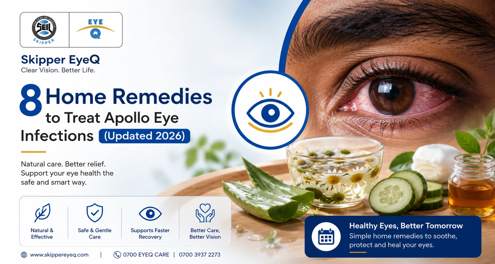
The 8 most effective home remedies for Apollo eye infection (conjunctivitis) are: (1) Saltwater rinse, (2) Warm compress, (3) Cold compress, (4) Raw honey eye wash, (5) Castor oil, (6) Green tea bags, (7) Colostrum for newborns, and (8) Tulsi (holy basil) water. These relieve symptoms only, they do not cure bacterial or severe viral infections. See a doctor if symptoms persist beyond 7 days.
Apollo eye infection, the common Nigerian name for viral conjunctivitis is one of the most prevalent and contagious eye conditions in West Africa, with outbreaks peaking during harmattan season and school terms. Most cases are viral and self-limiting, but the redness, discharge, and discomfort can be deeply uncomfortable. This guide covers 8 evidence-informed home remedies that can help manage symptoms, along with clear guidance on when home care is enough and when you need to see an eye specialist at Skipper Eye-Q.
For a full clinical overview of the condition itself, see our dedicated guide on how to cure Apollo eye disease and our page on the treatment of Apollo eye disease by type (viral, bacterial, allergic).
Apollo eye infection is the popular Nigerian and West African name for conjunctivitis inflammation of the conjunctiva, the transparent membrane covering the white of the eye and lining the inner eyelids. The name ‘Apollo’ comes from a 1969 epidemic of haemorrhagic conjunctivitis that coincided with the Apollo 11 moon landing.
| Type | Cause | Contagious? | Home Remedies Effective? |
|---|---|---|---|
| Viral (most common) | Adenovirus, enterovirus | YES — highly | YES — symptom relief |
| Bacterial | Staph, Strep, H. flu | YES | Partial — antibiotics needed |
| Allergic | Pollen, dust, dander | NO | YES — compresses, antihistamines |
| Irritant | Smoke, chemicals | NO | YES — rinse & compresses |
Important: Home remedies are most appropriate for viral and mild allergic conjunctivitis. Bacterial conjunctivitis often requires prescribed antibiotic eye drops. If you are unsure which type you have, read are your red eyes a sign of infection? before starting any home treatment.
Before starting home remedies, confirm your symptoms match Apollo/conjunctivitis rather than a more serious eye condition. See our full list of top common eye disorders and diseases to rule out other causes.
| Symptom | What It Looks Like | Indicates |
|---|---|---|
| Redness | Bloodshot whites, pink tinge | Inflammation of conjunctiva |
| Watery discharge | Clear, watery see | Viral conjunctivitis (most likely Apollo) |
| Yellow/green discharge | Thick pus, crusts overnight | Bacterial conjunctivitis, needs antibiotics |
| Itching | Persistent, worse with rubbing | Allergic conjunctivitis |
| Gritty sensation | Sand-in-eye feeling | Viral or bacterial infection |
| Light sensitivity | Squinting in normal light | Possible corneal involvement, see a doctor |
| Swollen eyelids | Puffy, tender lids | Moderate-to-severe infection |
If your eyes are watery without any other discharge, this may be allergic rather than infectious conjunctivitis, and antihistamines may work better than antibiotics or antimicrobial home remedies.
See our dedicated guide on tips to protect your eyes from conjunctivitis during monsoon for seasonal prevention strategies that work in the Nigerian climate.
Each remedy below includes: what it does, step-by-step instructions, safety notes, and evidence level. For a broader comparison of whether home remedies actually work for this condition, see our article home remedies for Apollo eye infections: do they work?.
Important safety note: All home remedies below are for symptom relief only. Never apply undiluted substances, urine, raw plant juices, or prescribed eye drops directly into your eye. If symptoms worsen after 48 hours of home treatment, stop and consult a doctor.
| Remedy | Best For | Evidence Level | Safety | Avoid If… |
|---|---|---|---|---|
| 1. Saltwater rinse | Discharge, crusting | Moderate | High | DIY concentration too strong |
| 2. Warm compress | Discharge, blepharitis, styes | High (AAO-backed) | High | Infection spreading |
| 3. Cold compress | Swelling, itching, redness | High | High | Severe pain or vision change |
| 4. Raw honey wash | Mild bacterial/viral | Low-moderate | Low-moderate | Allergy to bee products |
| 5. Castor oil | Dry, irritated eyes | Moderate | Moderate | Allergy to castor |
| 6. Green tea bags | Swelling, soothing | Moderate (anti-inflammatory) | High | Hot bags (burn risk) |
| 7. Colostrum (breast milk) | Neonatal conjunctivitis | Moderate (neonatal use) | High (newborns) | Adult infections |
| 8. Tulsi (holy basil) | Mild irritation | Low (traditional use) | Moderate | Direct eye contact without dilution |
Saline solution closely mimics the eye’s natural tears and has mild antimicrobial properties. It is effective at physically removing discharge, pus, and allergens from the eye surface, and is widely recommended as a first-line home measure for conjunctivitis symptom relief.
What it does: Mechanically clears discharge; antimicrobial properties reduce bacterial load on the eye surface
Evidence: Recommended by the American Academy of Ophthalmology as a supportive measure for conjunctivitis
Safety note: Never use table salt with iodine additive. Never apply hypertonic (overly salty) solution. Never use salt solution that has been stored overnight prepare fresh each time.
A warm compress is one of the most evidence-backed home remedies for eye infections and is explicitly recommended by the American Academy of Ophthalmology. It is particularly useful for loosening discharge that crusts the eyelids shut, and for relieving blepharitis that often accompanies recurrent eye infections.
What it does: Softens and loosens crusted discharge on eyelids; dilates meibomian glands to improve tear quality; soothes inflammation
Evidence: A 2014 clinical study of 22 participants showed warm compresses improve ocular surface health. Endorsed by AAO for infectious and non-infectious eye conditions.
Safety note: Do not use water that is uncomfortably hot. Do not press on the eyeball. If you notice the infection spreading to the other eye, stop and book a consultation at Skipper Eye-Q.
Where warm compresses help with discharge and gland function, cold compresses are better suited for the relief of itchy, inflamed, and swollen eyes particularly in allergic conjunctivitis or during the acute inflammatory phase of viral Apollo eye.
What it does: Constricts blood vessels to reduce redness and swelling; provides numbing relief from itching; reduces puffiness around the eyelids
Safety note: Never place ice directly on the eye or eyelid, it can cause ice burns to the delicate periocular skin. Do not press on the eye. Use the warm compress first if discharge and crusting are the main issue.
Warm vs Cold: When to use which?
Honey has well-documented antimicrobial, anti-inflammatory, and wound-healing properties. Research has shown that Manuka honey specifically can inhibit Staphylococcus aureus and Pseudomonas aeruginosa common bacterial causes of conjunctivitis. However, evidence for honey as a standalone treatment is still limited, and it should only be used as a supportive measure, not a replacement for prescribed antibiotics in bacterial cases.
What it does: Antimicrobial (inhibits bacterial growth); anti-inflammatory (reduces swelling and redness); creates a low-water environment unfavourable to bacteria
Safety note: Do not use commercially processed, sugared honey. Do not use undiluted honey directly in the eye. Do not use if you have a known bee product allergy. Stop immediately if burning or increased redness occurs.
Castor oil contains ricinoleic acid, an omega-9 fatty acid with documented anti-inflammatory properties. It is used in some lubricating eye drops as a base ingredient (notably Refresh Optive Advanced). As a home remedy, it can help with dry eye symptoms that worsen during an Apollo eye infection, and can reduce eyelid swelling when applied around (not inside) the eye.
What it does: Reduces periocular inflammation; lubricates the eyelid area; soothes itching associated with blepharitis
Safety note: Never instil castor oil directly inside the eye unless it is an ophthalmic-grade preparation. Keep away from the tear duct area. Do not use on children without medical supervision.
Green tea contains high concentrations of catechins (particularly EGCG) and tannins with documented anti-inflammatory and mild antimicrobial properties. Research published in the Journal of Agricultural and Food Chemistry demonstrated catechins from green tea can inhibit adenoviruses the primary cause of Apollo eye infection.
What it does: Reduces periocular swelling via vasoconstriction (tannins); anti-inflammatory catechins soothe irritated conjunctiva; provides a cooling, soothing physical sensation
Safety note: Never place warm or hot tea bags on the eye serious burn risk. Do not squeeze the bag to express tea directly into the eye. Chamomile tea bags are an alternative with similar anti-inflammatory properties.
Breast milk contains secretory IgA antibodies, lactoferrin, lysozyme, and epidermal growth factor — all of which have anti-infective properties. For neonatal conjunctivitis (ophthalmia neonatorum), small amounts of expressed breast milk have been used supportively in multiple studies. This remedy is specifically for newborns and infants; it is not recommended for adults.
What it does: Provides passive immunity via sIgA antibodies; lactoferrin binds iron needed by bacteria; lysozyme directly attacks bacterial cell walls
Safety note: Any newborn with eye discharge should be evaluated by a Skipper Eye-Q paediatric ophthalmologist immediately — neonatal conjunctivitis can be caused by gonorrhoea or chlamydia transmitted during birth, which require urgent systemic antibiotic treatment. Breast milk is supportive only and does not replace medical treatment.
See our children’s eye health and safety awareness guide for more on protecting newborn and infant eye health.
Tulsi (Ocimum sanctum) is a traditional Ayurvedic herb with documented antibacterial, antiviral, and anti-inflammatory properties. Laboratory studies have shown extracts of Ocimum sanctum inhibit Staphylococcus aureus and Streptococcus species — common bacterial causes of conjunctivitis. Clinical evidence in ophthalmology remains limited, so this remedy should be used as a supportive measure only.
What it does: Mild antimicrobial and anti-inflammatory properties; traditional use for conjunctivitis symptom relief
Safety note: Do not apply hot or warm tulsi water to the eye area. Do not apply undiluted tulsi juice or paste directly into the eye. If any irritation increases after application, stop immediately.
The following ‘remedies’ are commonly used in Nigeria and across West Africa for Apollo eye infection but have been shown to cause serious harm, including permanent vision loss. Skipper Eye-Q urges patients to avoid all of these:
| Harmful Remedy | Why People Use It | Why It Is Dangerous |
|---|---|---|
| Urine (human or animal) | Believed to be sterile and healing | Urine is NOT sterile; contains bacteria. Has caused severe corneal scarring and blindness. |
| Undiluted salt water | Perceived antimicrobial effect | Hypertonic solution damages corneal epithelium. Only use at prescribed dilution. |
| Soap / detergent | To ‘clean’ the eye | Causes severe chemical conjunctivitis; soap disrupts tear film |
| Lime / lemon juice | Vitamin C perceived as healing | Highly acidic; causes chemical burns to cornea |
| Petroleum jelly (Vaseline) | To soothe | Blocks meibomian glands; worsens infection |
| Raw onion juice | Traditional remedy | Highly irritating; can cause epithelial damage |
Viral Apollo eye infection typically lasts 7 to 14 days with or without home treatment. Home remedies shorten symptom severity but do not shorten the viral replication cycle.
For a detailed breakdown by infection type, see our full guide on how long Apollo eye infection lasts.
| Type | Without Treatment | With Home Remedies | With Medical Treatment |
|---|---|---|---|
| Viral (Apollo) | 7–14 days | 5–10 days (symptom relief only) | No antivirals typically; supportive only |
| Bacterial | Up to 2 weeks | Marginal improvement | 3–5 days with antibiotics |
| Allergic | Ongoing (until allergen removed) | Partial relief | Antihistamines most effective |
Apollo eye infection (viral conjunctivitis) is one of the most contagious conditions in routine clinical practice. A single infected household member can infect everyone in the home within 24 to 48 hours. See our precautions for viral conjunctivitis guide for a full prevention protocol, and our 10 tips for avoiding eye infections for year-round hygiene habits.
Do not attempt to manage Apollo eye infection at home if any of the following are present. Book an urgent consultation at Skipper Eye-Q.
Skipper Eye-Q operates specialist ophthalmology clinics at four locations across Lagos and Abuja. See all locations and opening hours.
Once you have recovered from an Apollo eye infection, the following measures reduce your risk of re-infection or spreading it to others during future outbreaks. See our complete prevention guide: how to prevent Apollo eye infection.
Apollo eye infection is uncomfortable but manageable at home in the majority of viral cases. The 8 remedies in this guide saltwater rinse, warm compress, cold compress, honey, castor oil, green tea bags, colostrum, and tulsi water are the most evidence-supported options available. Use them to relieve symptoms, practice strict hygiene to prevent spread, and stay informed about the warning signs that require professional care.
If you or a family member has an Apollo eye infection that is not improving after 7 days at home, book a consultation at Skipper Eye-Q. We have ophthalmologists available at four locations across Lagos and Abuja. Find your nearest branch.
There is no medical difference. ‘Apollo’ is the West African colloquial name for epidemic haemorrhagic conjunctivitis, popularised during a 1969 outbreak that coincided with the Apollo 11 moon landing. Medically, it is the same condition as viral conjunctivitis. For a full explanation, see our article on conjunctivitis: causes, symptoms and treatment.
No. Viral conjunctivitis cannot be cured in 24 hours the viral replication cycle takes time regardless of treatment. However, symptoms can be significantly reduced within 24 to 48 hours through consistent use of warm/cold compresses, saltwater rinses, and avoiding eye rubbing. For urgent relief options, see our guide on how long Apollo eye infection lasts.
No, undiluted tulsi juice or paste should never be applied directly to the eye. Only use cooled tulsi water as a compress or rinse around the eye. Direct instillation of undiluted botanical extracts can cause irritation and in some cases serious corneal damage.
Home remedies do not reliably kill bacteria causing conjunctivitis. Only antibiotic eye drops prescribed by a doctor can reliably eradicate bacterial conjunctivitis. For a complete guide to antibiotic options, see our article on antibiotics for pink eye. If you suspect bacterial Apollo eye (thick yellow-green discharge throughout the day), book a consultation at Skipper Eye-Q rather than attempting to treat it at home.
The safest approaches for children are warm compresses, saline rinses (properly diluted), and strict hygiene. Avoid honey, castor oil, tulsi, and essential oil-based remedies in young children without medical advice. See our children’s eye health guide for age-specific advice. If the child has thick discharge, pain, or significant light sensitivity, see a doctor immediately.
No. Remove contact lenses at the first sign of any eye infection and do not resume wearing them until all symptoms have completely resolved and you have completed any prescribed treatment. Book a comprehensive eye examination at Skipper Eye-Q before resuming lens wear after an infection.
No. You cannot contract Apollo eye infection by looking at an infected person. Transmission requires direct contact with infected eye secretions, touching contaminated surfaces then touching your own eye, or sharing personal items (towels, pillows, eye drops). Airborne transmission is not a route for conjunctivitis.
Have you ever noticed tiny spots or threads floating across your vision when looking at a bright sky or a plain wall? Or perhaps you've seen sudden flashes of light,...
Read More
If you're dealing with pain in your right eye along with a headache, it's most often one of a handful of familiar causes a migraine, a tension headache, a sinus...
Read More
Apollo, medically referred to as conjunctivitis occurs from inflammation of the eye's transparent membrane, known as the conjunctiva. This inflammation is triggered by infection or irritation caused by bacterial, viral,...
Read More