Cataract surgery is a common procedure that restores vision for millions of people worldwide. While the surgery itself is relatively straightforward, the recovery process requires careful attention to ensure optimal results. Among the many questions patients often ask, one of the most frequent is: “How long should I wait to wash my hair after cataract surgery?” This article explores this query in detail, providing insights into the recovery process and offering practical advice to help patients take the best care of their eyes. For expert cataract treatment, consider Skipper Eye-Q International Eye Hospitals, known for their advanced care and expertise.
Cataract surgery involves removing the clouded natural lens of the eye and replacing it with an artificial intraocular lens (IOL) to restore clear vision. The procedure, often conducted using advanced phacoemulsification techniques, is celebrated for its precision, minimal invasiveness, and efficiency. Typically completed within 20 to 30 minutes, it is one of the safest surgical interventions, with consistently high success rates.
Despite its straightforward nature, cataract surgery requires patients to exercise vigilance during the immediate postoperative period. The eye, rendered temporarily vulnerable due to the procedure, must be shielded from external contaminants and physical disruptions to facilitate healing. During this time, even seemingly mundane activities, such as washing hair, demand special attention and adaptation to avoid introducing bacteria or causing mechanical strain. By adhering to prescribed guidelines and prioritizing ocular safety, patients can support the recovery process and achieve the desired visual outcomes.
The timing and technique of hair washing following cataract surgery are crucial, but often patients overlook other daily routines and safety measures that are necessary for a full recovery. In the days and weeks after surgery, these extra care suggestions help promote healing, reduce discomfort, and preserve your eyesight.
Knowing when to wash your hair is crucial, but it’s just as critical to know how to do it comfortably and securely while you’re recovering. Minor technique modifications can avoid pressure on the recovering eye, infection risk, and unintentional discomfort.
Select the Safest Position for Hair Washing: Many patients prioritize timing above posture, which is crucial for safeguarding the operated eye. Some of the best and safest positions are:
A steady, comfortable posture helps prevent abrupt changes in the pressure surrounding the eye.
Choosing the Proper Shampoo Following Surgery: Even with care, hair products can inadvertently get into the eye. An additional degree of safety is added by using gentle products.
Comfort during the healing process can be maintained using easy-to-use, non-irritating products.
Controlling your anxiety during your first Hair wash: After cataract surgery, many people experience anxiety when washing their hair for the first time. This can be handled via:
After the first safe experience, most patients feel more confident and rapidly become used to the routine.
Indications of Too Early or Too Forceful Hair Washing: Some warning indicators suggest the eye may require additional protection even when precautions are taken:
If these happen, wait a few more days before washing your hair and see an ophthalmologist if the symptoms don’t go away.
Slow Restart of Regular Hair Care Practices: Following cataract surgery, recovery is gradual. Gradually, hair washing precautions might be relaxed:
A progressive approach helps to avoid recovery setbacks.
Surgical approach, healing reaction, and general eye health all affect recovery times. Although broad recommendations are useful, specific instructions are usually given precedence. In order to guarantee safe hygiene habits and the best possible healing results, patients recovering under the supervision of doctors at Skipper EyeQ are usually given personalized assistance.
Our ophthalmologists generally advise for a waiting period of 24 to 48 hours before hair washing. This interval facilitates the establishment of an initial protective barrier over the surgical site, mitigating the risk of microbial contamination or mechanical disruption from water or cleaning agents. Nonetheless, individual variations in recovery and specific procedural nuances underscore the importance of adhering to surgeon-specific recommendations.
The primary concern associated with premature hair washing is inadvertent exposure of the surgical eye to waterborne irritants or pathogens. Even trace amounts of water may compromise the sterility of the healing ocular surface, posing a heightened risk of endophthalmitis or delayed epithelialization.
Several variables mediate the appropriate timing for resuming hair washing post-surgery:
Advanced methods, such as micro-incisional phacoemulsification, tend to expedite healing due to minimized tissue disruption. Conversely, surgeries requiring larger incisions or addressing complex ocular pathologies may necessitate prolonged recovery intervals.
Recovery rates exhibit inter-individual variability, influenced by factors such as age, systemic health status, and compliance with post-operative care protocols. For example, patients with diabetes or immunocompromised conditions may experience protracted healing timelines.
Customized guidance from the operating surgeon reflects an integration of procedural specifics, intraoperative findings, and individual patient risk profiles. Deviation from generalized timelines should be predicated on such expert advice.
Living in environments characterized by elevated humidity, dust, or pollution necessitates heightened vigilance to shield the vulnerable eye from exogenous irritants during the recovery period.
Upon receiving clearance from your ophthalmologist, resuming hair washing should proceed with meticulous care. A stepwise strategy is outlined below:
Ensure compliance with the specified timeframe, which typically ranges from 24 to 48 hours. If uncertainties persist, seek clarification from our expert team of ophthalmologists.
Employ protective eyewear, such as a shield or goggles, to preclude water, shampoo, or soap from contacting the surgical eye.
Select hypoallergenic and tear-free shampoos to minimize the risk of chemical irritation. Avoid fragranced or harsh formulations that could exacerbate discomfort in case of inadvertent exposure.
Regulate the water stream to a gentle flow and maintain a lukewarm temperature. High-pressure sprays or excessively hot water should be avoided due to their potential to dislodge protective ocular barriers or cause irritation.
Tilt your head backward to direct water away from your face and eyes. This posture minimizes the likelihood of accidental contact with the healing eye.
Even beyond the immediate post-operative period, maintaining vigilance during hair washing is essential for optimal recovery after cataract surgery. Adhering to these precautions can significantly enhance healing outcomes:
Refrain from rubbing, touching, or applying pressure near the surgical eye. Mechanical irritation can disrupt the healing process and increase the risk of infection or complications.
Hot water and steam can exacerbate ocular inflammation, dilating blood vessels and promoting swelling. Additionally, these environments may encourage microbial growth, increasing the risk of post-surgical infections. Stick to lukewarm water to mitigate these risks.
Steer clear of using hair care products such as conditioners, serums, or sprays during the early recovery period. These products may inadvertently come into contact with the eye, causing irritation or introducing contaminants to the healing area.
For patients unsure about safely washing their hair while protecting the eye, enlisting the help of a caregiver can provide additional security. This ensures that protective measures, such as shielding the eye, are effectively implemented.
Remain vigilant for any signs of complications post-washing, such as persistent redness, swelling, increased discomfort, or unusual discharge. If any of these symptoms occur, promptly consult our ophthalmologists for quick evaluation and guidance.
By incorporating these precautions into your post-operative routine, you can ensure a safer recovery and support the restoration of optimal visual health.
Hair washing, while seemingly mundane, acquires heightened significance in the context of cataract surgery recovery. By adhering to the recommended waiting period, employing appropriate protective measures, and diligently observing post-operative guidelines, patients can safeguard their surgical outcomes. Ultimately, patient-specific adaptations, guided by clinical expertise, remain paramount in ensuring a seamless recovery and the restoration of optimal visual function. For expert cataract care and recovery guidance, consider Skipper Eye-Q International Eye Hospitals, renowned for their comprehensive and patient-centric approach to ophthalmology.
After cataract surgery, your eye is still healing and is very sensitive. So, if you get shampoo or soap into your eyes, it will cause burning. Moreover, there can be redness and watering, and there are chances of infection as well. This is because when your eyes are healing, the chemicals in the shampoo can be too harsh.
Furthermore, in Nigeria, the weather can be hot as well as dusty. So, your eyes can feel dry or irritated after the surgery. In this case, if shampoo gets into your eyes, do not rub them. Instead, you need to wipe it off gently around the closed eye. Make sure to use a clean, wet cloth.
As per the doctor’s advice, you should at least wait for one to two weeks before letting water touch your eyes directly. Moreover, be very careful during this time and bathe or wash your face with maximum care. Also, avoid swimming pools, rivers, or lakes. It is because these places carry a lot of germs, so it increases your chances of getting an infected eye.
After about two weeks, you can carefully splash some clean and lukewarm water on your closed eyes. However, you need to avoid rubbing or pressing. As Nigeria’s climate can sometimes make your eyes sweaty or dusty, you can gently pat your face dry instead of washing it too soon.
No, it is strictly forbidden to rub your eyes after cataract surgery. It is because if you rub your eyes, it can move the new lens. Moreover, there is an increased chance of infection as well. Now, in Nigeria, dust and heat can make your eyes feel itchy or uncomfortable. So, instead of rubbing, you can use special eye drops that are given by your doctor. These drips will reduce the itching and keep your eyes moist.
Url – https://skippereyeq.com/home-remedies-for-itchy-eyes-or-dry-eyes/


Quick Answer
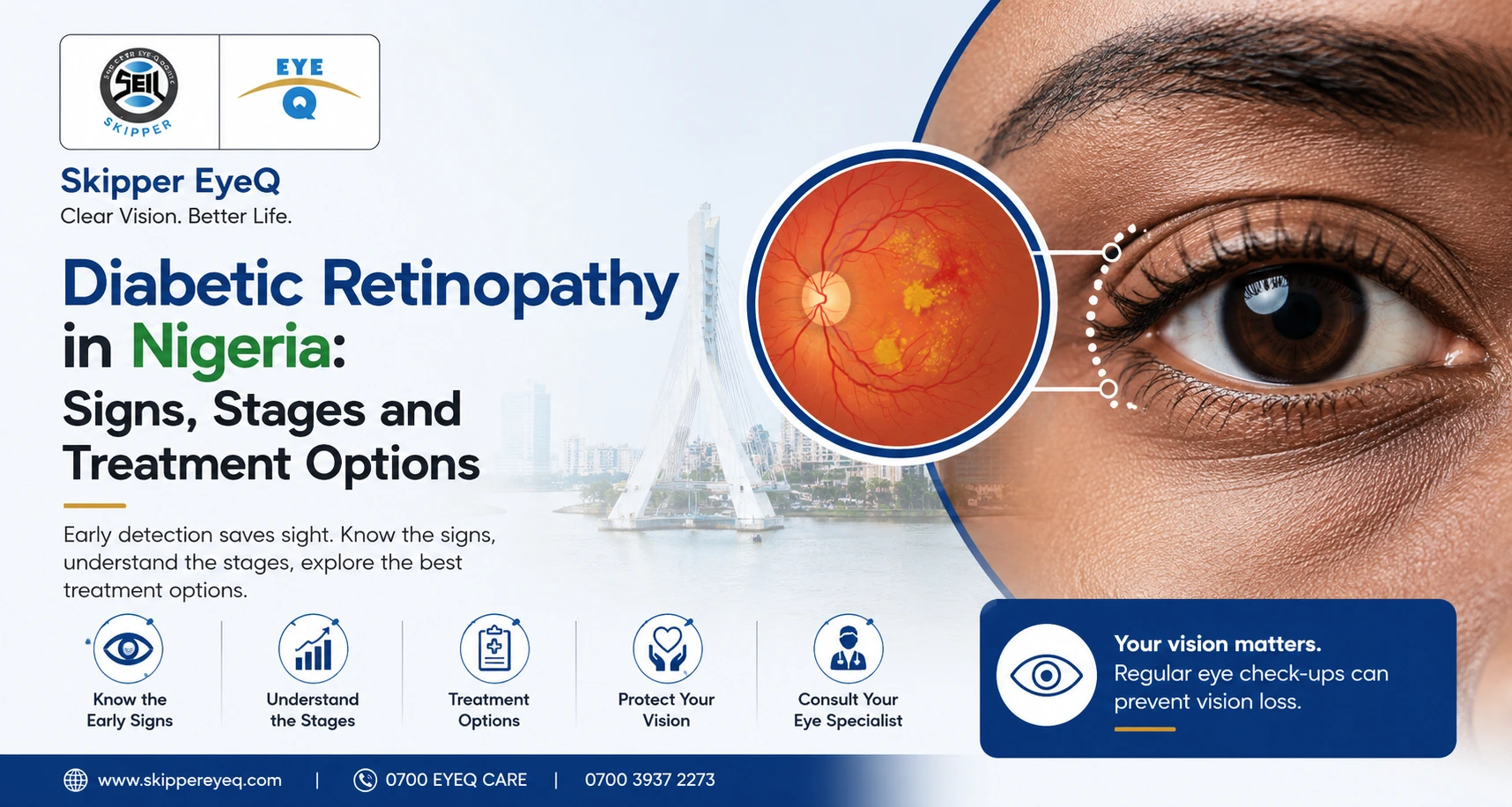
Diabetic retinopathy is an eye disease caused by high blood sugar damaging the tiny blood vessels at the back of the eye. It is the leading cause of...
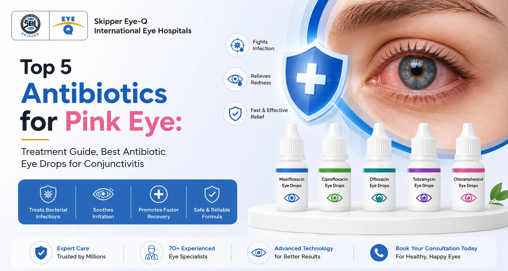
The top 5 antibiotics for bacterial pink eye are: (1) Tobramycin eye drops, (2) Erythromycin ointment, (3) Ciprofloxacin eye drops, (4) Ofloxacin eye drops, and (5) Gentamicin eye drops. These...
Read More
Quick Answer
Antallerge eye drop is a sterile eye medicine used in Nigeria to relieve red, itchy and watery eyes caused by allergies. It contains two ingredients antazoline (which stops...

Laser eye surgery is a technologically advanced procedure. Through laser eye surgery, it provides permanent correction to all the problems relating to vision; the issues involve nearsightedness, farsightedness, and astigmatism. However, a big question remains- whether the whole process is painful. Is laser eye surgery painful? This is often asked, and knowing the level of discomfort before, during, and after the procedure may or may not clear up some anxiety. At Skipper Eye-Q Super Speciality Eye Hospital, we address these concerns and are dedicated to providing safe, effective vision correction through state-of-the-art laser technology. With more than 5 million successful surgeries in the past 15 years, we have a track record of a 100% success ratio.
This article digs out the things that influence pain in surgery, some tips on managing pain, and home remedies for any discomfort felt after laser eye treatment.
One of the most significant fears among people when they hear about laser eye surgery is the pain that they will face. Although it should be one of the major concerns, it is not the reality because most patients claim to have suffered very minimal discomfort in the process both during and after the procedure. Is laser eye surgery painful? The latter depends on individual pain tolerance and the specific technique used.
In fact, the whole procedure is quite painless with local anesthetic drops in your eye. The droplet itself numbs the surface of your eye, meaning that you never feel a sudden pain while performing the treatment. The laser surgery itself is painless though some patients complain of pressure-like sensations or may feel uncomfortable when it reshapes their cornea; however, such sensations are always brief and are not considered to be painful.
Generally, after the surgery, one might feel minimal discomfort and sometimes dry eyes. These conditions are temporarily faced and are very easy to manage. The degree of pain varies from patient to patient. It mainly depends on the other patients who have faced this surgery. For most patients, it can last for hours and even up to a couple of days at maximum. This is one of the reasons why advanced technology has produced the best laser eye surgery techniques, ensuring there is maximum comfort for the patient.
There are many factors that influence a patient’s pain perception in laser eye surgery. Below, we have stated some of the key ones.
The intensity of pain also depends on the kind of laser eye surgery you are undergoing. For example, LASIK is one of the most common procedures wherein a small flap of your cornea is made to allow the laser to reshape your eye’s surface. “Patients usually go through little or no pain during LASIK, and the discomfort only sometimes comes after its successful performance”, says a surgeon.
The other form of laser surgery is PRK, where the outer layer of the cornea is typically scraped off, and it hurts more and is more uncomfortable when healing. However, these are very effective procedures, although LASIK tends to be even more effective than that with recovery speed and the reduction of the level of pain involved.
Each individual has pain tolerance. Therefore, some may be sore, while for others this could be much more distracting. However, laser eye surgery is designed to be as minimally uncomfortable as needed for the most part. You can speak about your pain level during your first laser eye surgery consultation so that the ophthalmologist can plan the procedure bearing your pain thresholds in mind.
The expertise of your surgeon and the technology used also determine your experience during laser eye surgery. Advanced equipment and experienced surgeons reduce the risks associated with complications and discomfort. A good clinic that delivers the best in laser eye surgery consultation usually has the latest technology available for better accuracy and comfort of the procedure. With a team of highly trained eye specialists and advanced infrastructure, Skipper Eye-Q Super Speciality Eye Hospital stands as one of the most reliable eye hospitals. Our ophthalmologists have decades of experience in cataracts, retina, glaucoma, presbyopia, cornea, squint, and refractive surgeries.
There may be a few eye conditions that can be a nuisance after your surgery, for example, dry eyes. Be certain to discuss any pre-existing conditions during the consultation, so your surgeon can take any necessary precautions.
The post-operative care will greatly influence the outcome of healing and the extent of pain or discomfort you will endure. Strict compliance with the eye drops, avoidance of strenuous activities, and strict compliance with the surgeon’s recommendation regarding rest for your eyes will greatly help minimize discomfort. Non-adherence to the above may result in complications, hence increased chances of more pain or irritation.
Despite the painlessness of laser eye surgery, some guidelines help manage pain if it arises. Here are some effective tips that help minimize discomfort both during and after the procedure:
A reliable surgeon will provide you with complete post-operative care guidelines that detail eye care, medication use, and application of eye drops. All these steps can help avoid infections and minimize pain and discomfort after a laser eye treatment, ensuring optimal healing. At Skipper Eye-Q, we ensure the smooth recovery of patients by providing them with comprehensive post-surgery care.
Dry eyes are one of the most common complaints after laser eye surgery. Your ophthalmologist will prescribe lubricating eye drops to help manage this. These drops help keep the eyes moist and reduce dryness and irritation. However, patients should use them as prescribed by their ophthalmologist to reduce discomfort effectively.
After laser eye surgery, your eyes may be more sensitive to light. Wearing sunglasses helps protect your eyes from glare and bright lights, which can reduce the chance of discomfort. It’s particularly useful when you’re outdoors, especially in the first few days following surgery.
Avoid intense visual focus activities such as reading or working on a computer after surgery. This will allow your eyes to rest and recover and reduce irritation and discomfort. Be sure to follow your surgeon’s guidelines on when to resume normal activities.
For some minor discomforts, your physician may recommend ibuprofen or acetaminophen over the counter. It will help soothe any ache, but seek your ophthalmologist’s counsel on medication post-surgery for pain.
Rehydration is part of recovery. Drink plenty of water to keep your body hydrated, healing and reducing the risk of dry eyes on or after surgery. Good hydration helps ensure that your eyes are healthy, and it even helps minimize the side effects of the procedure.
In the first days after laser eye treatment, screen time should be minimized. Too much digital screen time can exacerbate dryness and strain on your eyes. Stick to your surgeon’s advice about screen time so that your eyes can heal appropriately.
Dry air also causes irritation after the surgery of your laser eyes if you spend more hours in your rooms because of having been set your heaters or coolers. Moistening the room air by putting up a humidifier will retain moist air, ensuring that you have not developed dried-out eyes due to discomfort.
Swimming in pools, lakes, and the ocean is not advised to be done in the healing time of your eyes. Chlorine combined with the bacterial impact of the water will cause irritation in the eyes and promote infection. One should wait for an ophthalmologist’s confirmation after a laser eye surgery consultation so that the specialist allows them to resume swimming.
While professional medical care is useful right after laser eye surgery, many home remedies could help you relieve mild discomfort felt after the operation. These treatments can complement your recovery process to make you feel better.
Apply a cold compress to help reduce swelling and discomfort. Apply a clean, cold compress over closed eyelids to soothe irritation or dryness, a common condition following surgery. Make sure to follow your eye specialist’s advice about the frequency of application.
The time your eyes have to rest during this period of surgery is important in recovery. Try avoiding things that exert more strain on the eyes, including using electronic gadgets or reading long stretches. Also, try sleeping with your eyes closed, giving it time to rest.
The most common feeling after laser eye surgery is the dryness and irritation of the eyes. Artificial tears can sometimes be quite helpful in removing this condition. These artificial tears are readily available in any over-the-counter drug store. Patients can apply these artificial tears as frequently as necessary during the day. However, consult your ophthalmologist for the dosage and frequency of usage.
While it might be tempting to rub your eyes if they feel irritated, it’s essential to avoid doing so after surgery. Rubbing your eyes can disrupt the healing process and introduce bacteria, which may lead to infection. Always follow your surgeon’s instructions for proper eye care.
Use a humidifier in your home. This will ensure that the humidity in the air will keep the moisture within, preventing your eyes from becoming dry. This dry environment will not help with your comfort following the laser eye surgery, and keeping the environment moist can also help with comfort and healing.
Most laser eye surgery is pain-free; hence most patients do not find it troublesome to undergo it. You would sometimes feel the feeling of dryness or minor irritation after a surgical procedure; this is normally for a while, and proper care usually takes it away. Thus, for patients in need of an irreversible method to correct vision impairments, the best laser eye surgery comes in as an easy, harmless technique that restores vision and results in a greater quality of life.
At Skipper Eye-Q Super Speciality Eye Hospital, comfort and safety are guaranteed at every step of your laser eye treatment. Our finest laser technology is accompanied by a patient-first approach, which ensures a smooth transition from consultation to recovery and beyond the procedure. We provide complete post-operative eye care services to make your experience as comfortable and convenient as possible for you!
Quick Answer
Diabetic retinopathy is an eye disease caused by high blood sugar damaging the tiny blood vessels at the back of the eye. It is the leading cause of...
The top 5 antibiotics for bacterial pink eye are: (1) Tobramycin eye drops, (2) Erythromycin ointment, (3) Ciprofloxacin eye drops, (4) Ofloxacin eye drops, and (5) Gentamicin eye drops. These...
Read More
Quick Answer
Antallerge eye drop is a sterile eye medicine used in Nigeria to relieve red, itchy and watery eyes caused by allergies. It contains two ingredients antazoline (which stops...

Glaucoma is a serious eye condition that can lead to permanent vision loss if left untreated. It is often referred to as the “silent thief of sight” because it progresses slowly and without noticeable symptoms in the early stages. Understanding this condition can help you recognize the warning signs and seek timely treatment. Consult the expert at Skipper Eye Q and learn more about it.
Glaucoma is not a single disease but a group of eye conditions that damage the optic nerve, which is responsible for transmitting visual information from your eyes to your brain. This damage is often caused by high intraocular pressure (pressure inside the eye). However, it’s important to note that glaucoma can also occur even with normal eye pressure.
The condition can affect people of all ages, but it is more common in older adults. Early detection and management are crucial to prevent irreversible vision loss.
Glaucoma has several causes, which can vary based on its type. Here are the common factors:
The most common type of glaucoma occurs when the drainage system in the eye becomes less efficient, leading to a gradual build-up of pressure.
In this type, the angle between the iris and cornea becomes blocked, causing a rapid increase in eye pressure.
Family history plays a significant role. If someone in your family has glaucoma, your chances of developing it are higher.
Eye injuries, surgeries, or severe infections can damage the optic nerve or affect eye pressure, increasing the risk of glaucoma.
Conditions like diabetes, hypertension, and certain eye disorders can contribute to the development of glaucoma.
Long-term use of steroid medications can increase eye pressure, leading to secondary glaucoma.
In its early stages, glaucoma often shows no symptoms. This is why regular eye check-ups are crucial, especially if you fall into the high-risk category. However, as the condition progresses, you may notice the following signs:
This is the most common type. Symptoms include:
This type is less common. Symptoms include:
Symptoms include:
There are several different types of glaucoma, each with its own unique characteristics and causes. Here are some of the most common types:-
Regular eye examinations are the best way to catch glaucoma early. During a comprehensive eye check-up, your ophthalmologist may perform the following tests:
The goal of glaucoma treatment is to reduce eye pressure and prevent further optic nerve damage. While vision already lost cannot be restored, timely treatment can protect your remaining eyesight.
The first step in treating glaucoma often involves medications to help manage eye pressure.
When medications are not sufficient, laser therapy can be an effective treatment option to treat glaucoma.
When medications and laser treatments are not enough, surgical procedures may be recommended:
Making some changes in your daily habits can significantly aid in managing glaucoma and maintaining overall eye health. Here are some essential lifestyle modifications to consider:
While you cannot completely prevent glaucoma, you can reduce the risk of severe damage by following these steps:
Being diagnosed with glaucoma can be overwhelming, but with proper management, you can lead a normal life. Regular follow-ups with your ophthalmologist are essential to monitor the condition. Make sure to adhere to your treatment plan and report any changes in your vision immediately.
Support groups or online communities can provide emotional support and practical tips for managing daily challenges.
If you experience any of the following symptoms, it’s crucial to seek medical attention right away:
Glaucoma is a serious condition that requires timely detection and consistent care. While it cannot always be prevented, early diagnosis and treatment can help preserve your vision. Regular eye check-ups, especially after the age of 40 or if you have risk factors, are key to protecting your eyesight.
By staying vigilant about your eye health and following medical advice, you can reduce the impact of glaucoma and maintain a good quality of life. If you suspect any issues with your vision, don’t wait—consult an eye specialist promptly.
Quick Answer
Diabetic retinopathy is an eye disease caused by high blood sugar damaging the tiny blood vessels at the back of the eye. It is the leading cause of...
The top 5 antibiotics for bacterial pink eye are: (1) Tobramycin eye drops, (2) Erythromycin ointment, (3) Ciprofloxacin eye drops, (4) Ofloxacin eye drops, and (5) Gentamicin eye drops. These...
Read More
Quick Answer
Antallerge eye drop is a sterile eye medicine used in Nigeria to relieve red, itchy and watery eyes caused by allergies. It contains two ingredients antazoline (which stops...

Retinal detachment is a serious eye condition that can lead to vision loss if not treated promptly. It occurs when the retina, the light-sensitive layer at the back of the eye, separates from its underlying supportive tissue. This blog aims to explain the symptoms, diagnosis, and treatment options for retinal detachment in a way that is easy to understand, without using medical jargon.
To put it simply, the retina is like the film in a camera. It captures the images you see and sends them to the brain via the optic nerve. When the retina detaches, it loses its connection to the blood vessels that supply it with oxygen and nutrients, leading to a risk of permanent vision loss.
There are three main types of retinal detachment:
Retinal detachment can affect anyone, but some people are at higher risk due to certain factors. These include:
Recognizing the symptoms of retinal detachment early can be crucial in saving your vision. The symptoms often appear suddenly and include:
If you experience any of these symptoms, it’s important to seek medical attention immediately. Delaying treatment can lead to permanent vision loss.
Diagnosing retinal detachment involves a thorough examination of the eye by an ophthalmologist. Here’s what you can expect during the diagnosis process:
These diagnostic tools help the doctor determine the location and extent of the detachment, which is essential for planning the right treatment.
The goal of treatment is to reattach the retina and restore vision as much as possible. The treatment depends on the type, size, and location of the detachment. Here are the most common treatment options:
A laser beam is directed at the retina through the pupil. The laser creates tiny burns around the retinal tear, causing scarring that seals the retina to the underlying tissue. This method is often used for small tears or holes in the retina before they lead to a full detachment.
The doctor applies a freezing probe to the outer surface of the eye. The freezing process causes a scar to form, which helps secure the retina in place. Cryopexy is often used in combination with other procedures to treat tears that could lead to detachment.
A gas bubble is injected into the eye’s vitreous cavity. The bubble presses against the detached retina, pushing it back into place. Laser or freezing treatment is then used to seal the tear. This method is effective for certain types of detachments, particularly those in the upper part of the retina.
A tiny, flexible band (scleral buckle) is placed around the outside of the eye. This band gently pushes the eye inward, allowing the retina to reattach to the eye’s wall. The band remains in place permanently. This is often accompanied by other procedures like laser therapy or cryotherapy. Scleral buckling is commonly used for rhegmatogenous detachment. It provides long-term support to keep the retina attached.
The vitreous gel that is pulling on the retina is removed and replaced with a gas bubble, silicone oil, or saline. This procedure is often combined with laser or freezing treatments to secure the retina. Vitrectomy is usually reserved for more severe or complicated detachments, including those associated with diabetic retinopathy.
Recovery from retinal detachment treatment varies depending on the procedure performed and the extent of the detachment. Here’s what you can generally expect during the recovery period:
While you can’t always prevent retinal detachment, you can reduce your risk by taking certain precautions:
Even after successful treatment, life with retinal detachment may require some adjustments. Here are a few tips to help you manage:
Here are common early signs:
If you experience any of the above symptoms, it’s crucial to see an ophthalmologist immediately. A detached retina is a medical emergency, and prompt treatment is essential to prevent permanent vision loss. Here are some steps to take:
Remember, early detection and treatment are key to preserving your vision. If you have any concerns about your eye health, don’t hesitate to reach out to a medical professional.
Retinal detachment is a serious condition, but with early detection and appropriate treatment, many people can retain their vision. Being aware of the symptoms and understanding the treatment options can make a significant difference in the outcome. Regular eye exams and protecting your eyes from injury are key preventive measures. If you ever notice symptoms like flashes of light, floaters, or a shadow in your vision, don’t hesitate to seek immediate medical attention. For expert care, visit Skipper Eye-Q International Eye Hospital. Their specialists are ready to help you preserve your vision with advanced treatments. Schedule a consultation today.
Quick Answer
Diabetic retinopathy is an eye disease caused by high blood sugar damaging the tiny blood vessels at the back of the eye. It is the leading cause of...
The top 5 antibiotics for bacterial pink eye are: (1) Tobramycin eye drops, (2) Erythromycin ointment, (3) Ciprofloxacin eye drops, (4) Ofloxacin eye drops, and (5) Gentamicin eye drops. These...
Read More
Quick Answer
Antallerge eye drop is a sterile eye medicine used in Nigeria to relieve red, itchy and watery eyes caused by allergies. It contains two ingredients antazoline (which stops...

When we talk about glaucoma, the conversation usually revolves around the more common forms such as primary open-angle glaucoma or angle-closure glaucoma. However, there’s a lesser-known but equally important type called ghost cell glaucoma. This condition can be quite serious, particularly because it often follows complications from eye trauma or surgery. Understanding ghost cell glaucoma is crucial, not just for those diagnosed with it, but for anyone interested in maintaining good eye health and preventing vision loss.
In this comprehensive blog, we’ll explore ghost cell glaucoma in detail, from its causes and symptoms to its diagnosis and treatment.
Ghost cell glaucoma is a secondary form of glaucoma that arises due to the presence of “ghost cells” in the eye. These ghost cells are altered red blood cells that have undergone significant changes after bleeding in the anterior chamber of the eye (the front part). Normally, the aqueous humor, a clear fluid inside the eye, flows out through the drainage system. However, when ghost cells obstruct this drainage system, intraocular pressure (IOP) rises, leading to damage of the optic nerve—a condition known as glaucoma.
Several factors can lead to ghost cell glaucoma:
Ghost cell glaucoma shares some symptoms with other types of glaucoma, making it essential to pay close attention to changes in your eye health. Here’s what you should look out for:
If you experience any of these symptoms, it’s crucial to see an eye doctor right away.
Getting an accurate diagnosis is crucial for effective management. Here’s how your eye doctor might approach diagnosing ghost cell glaucoma:
Treatment for ghost cell glaucoma focuses on reducing intraocular pressure and addressing the underlying cause of the condition. The specific treatment approach will depend on the severity of the glaucoma and the individual patient’s needs. Here are some common treatment options:
The initial approach to treating ghost cell glaucoma focuses on reducing intraocular pressure through non-invasive methods. Medical management primarily involves:
Eye drops are usually the first step in managing ghost cell glaucoma. These medications help reduce intraocular pressure (IOP) by either decreasing the production of fluid in the eye or increasing its drainage. Commonly prescribed eye drops include:
In some cases, oral medications may be prescribed to further reduce IOP. These are typically used when eye drops alone are not sufficient.
Several surgical options are available depending on the severity of the condition:
In this procedure, a small incision is made in the cornea, and a balanced salt solution (BSS) is used to flush out the ghost cells from the anterior chamber. This helps reduce intraocular pressure (IOP) by clearing the trabecular meshwork.
Trabeculectomy involves creating a new drainage pathway for the aqueous humor. A partial-thickness flap is made in the sclera, and an opening is created in the trabecular meshwork, allowing the aqueous humor to drain into the subconjunctival space, forming a bleb that regulates the pressure.
This technique is used to reduce intraocular pressure by directly draining the aqueous humor. A needle punctures the anterior chamber, allowing the fluid to flow out. It is a temporary measure for rapid IOP reduction and can be repeated if necessary.
Glaucoma drainage devices, such as tube shunts, provide an alternative pathway for aqueous humor drainage, bypassing the blocked trabecular meshwork. These devices help maintain aqueous outflow and control intraocular pressure.
Laser trabeculoplasty uses a laser to create small burns on the trabecular meshwork, leading to remodeling and improved drainage. Although commonly used in open-angle glaucoma, it can be considered in cases where ghost cells cause persistent blockages.
Cyclodestructive procedures, such as cyclophotocoagulation or cyclocryotherapy, reduce the production of aqueous humor by partially destroying the ciliary body. These methods are generally reserved for cases where other treatments have failed or are unsuitable.
A vitrectomy is sometimes necessary if there is significant vitreous hemorrhage contributing to ghost cell glaucoma. During this procedure, the vitreous gel (containing the ghost cells) is removed from the eye and replaced with a clear solution, helping to restore normal fluid dynamics and reduce IOP.
Each of these interventions is chosen based on the patient’s response to initial treatments.
Regular follow-up visits are essential to monitor IOP and adjust treatment as needed. Patients with ghost cell glaucoma need ongoing care to prevent further vision loss. During these visits, the eye doctor will assess the effectiveness of the treatment, check for any changes in vision, and ensure that IOP remains under control.
Along with medical and surgical treatments, certain lifestyle changes can help manage ghost cell glaucoma more effectively:
While not all cases can be prevented, there are several measures you can take to reduce your risk:
Coping with ghost cell glaucoma requires a proactive approach and a strong support system. Here are some practical tips for managing your condition:
Ghost cell glaucoma may not be as commonly discussed as other forms of glaucoma, but it is a significant condition that warrants attention. By understanding its causes, symptoms, and treatment options, you can take proactive steps to protect your vision and maintain your eye health. Regular eye exams, timely treatment, and lifestyle adjustments are key to managing this condition effectively.
If you suspect you have ghost cell glaucoma or are experiencing related symptoms, seek medical advice promptly. Early intervention can make a significant difference in preserving your vision and ensuring a better quality of life. Remember, taking care of your eyes is not just about treating symptoms but about maintaining your overall well-being and quality of life.
Quick Answer
Diabetic retinopathy is an eye disease caused by high blood sugar damaging the tiny blood vessels at the back of the eye. It is the leading cause of...
The top 5 antibiotics for bacterial pink eye are: (1) Tobramycin eye drops, (2) Erythromycin ointment, (3) Ciprofloxacin eye drops, (4) Ofloxacin eye drops, and (5) Gentamicin eye drops. These...
Read More
Quick Answer
Antallerge eye drop is a sterile eye medicine used in Nigeria to relieve red, itchy and watery eyes caused by allergies. It contains two ingredients antazoline (which stops...

Have you ever wondered about the potential risks of cataract surgery? While this procedure is generally safe and effective in restoring vision, like any surgical intervention, it comes with potential complications. Understanding these risks and knowing how to manage them can significantly contribute to a successful recovery. In this blog, we will explore the five most common complications of cataract surgery and practical tips on dealing with them.
Cataract surgery is a widely performed procedure aimed at restoring clear vision by removing the clouded natural lens of the eye and replacing it with an artificial intraocular lens (IOL). The surgery is typically quick and performed under local anesthesia, and most patients experience significant improvement in their vision within a few days. The primary goal is to help patients regain their visual clarity and improve their quality of life.
During the surgery, the ophthalmologist makes a small incision in the eye, uses ultrasound waves to break up the cloudy lens, and then gently removes it. The new artificial lens is then placed into the same position. The procedure generally takes less than 30 minutes and is known for its high success rate.
However, as with any surgical procedure, there are certain risks and complications associated with cataract surgery. It is essential to be aware of these potential issues and understand that most complications can be effectively managed with prompt medical attention and care.
Post-surgical infection is a potential complication following cataract surgery. It occurs when harmful bacteria or pathogens enter the eye during or after the procedure. Common causes include insufficient sterilization of surgical instruments, contamination during the surgery, or improper post-operative care. Though rare, an infection can seriously affect the eye’s healing process and overall health.
Patients should be aware of symptoms indicating an infection. These include increased redness in the eye, persistent pain, unusual discharge, or blurred vision that worsens instead of improving over time. If any of these symptoms are noticed, it is essential to seek medical attention promptly.
Treating an infection typically involves using antibiotic eye drops or oral antibiotics, prescribed by your ophthalmologist. In some cases, additional interventions may be required to manage the infection effectively.
Avoid touching or rubbing your eyes, especially with unwashed hands. Follow all post-operative care instructions provided by your healthcare team, and attend scheduled follow-up appointments to monitor your eye’s healing process. Maintaining good hygiene and adhering to medical advice can significantly reduce the risk of post-surgical infections.
Edema, or swelling, is a common occurrence in the eye tissues following cataract surgery. This swelling is usually temporary and part of the body’s natural healing process. It can cause discomfort and blurred vision during the recovery period but typically resolves with proper care and time.
Swelling after cataract surgery can be caused by several factors. The body’s response to the surgical procedure itself often leads to inflammation and fluid accumulation. Other contributing factors can include prolonged surgery time, pre-existing conditions like diabetes, or the use of certain medications. Physical strain and overactivity shortly after the operation can also exacerbate swelling.
Managing edema involves several practical steps. Applying cold compresses to the closed eye can help reduce swelling and provide relief. It is crucial to follow your doctor’s advice on activity levels, avoiding strenuous activities that could strain the eye. Your ophthalmologist may prescribe anti-inflammatory eye drops to help alleviate the swelling.
Rest is equally important, allowing your body to heal. Maintaining a healthy diet rich in vitamins and minerals can also support the healing process. Regular follow-up appointments with your doctor will ensure that the swelling is monitored and managed effectively, contributing to a smooth recovery.
Retinal detachment is a serious condition where the retina, the light-sensitive tissue at the back of the eye, separates from its underlying supportive tissue. This detachment disrupts the retina’s function and can lead to permanent vision loss if not treated promptly. Although rare, retinal detachment can occur as a complication following cataract surgery.
Several factors can increase the risk of retinal detachment after cataract surgery. These include severe nearsightedness (myopia), a history of retinal detachment in the other eye, previous eye injuries, and certain eye conditions like lattice degeneration. Individuals who have undergone multiple eye surgeries are also at higher risk.
Recognizing the early signs of retinal detachment is crucial for prompt treatment. Symptoms to watch for include sudden flashes of light, an increase in floaters (tiny specks that drift through your field of vision), and the appearance of a dark shadow or curtain over part of your visual field. These symptoms warrant immediate medical attention.
If retinal detachment occurs, it requires urgent surgical intervention to reattach the retina and restore vision. Common procedures include laser surgery, cryopexy (freezing), or pneumatic retinopexy, where a gas bubble is injected into the eye to push the retina back into place. In more severe cases, a vitrectomy, which involves removing the gel-like substance in the eye, may be necessary.
A secondary cataract, also known as posterior capsule opacification (PCO), is a common complication that can occur after cataract surgery. This happens when the back of the lens capsule, which holds the artificial lens in place, becomes cloudy over time, leading to a decrease in vision. It’s important to note that this is not a true cataract but rather a side effect of the surgery.
Symptoms of a secondary cataract can include blurred or cloudy vision, glare sensitivity, difficulty reading or recognizing faces, and experiencing halos around lights. These symptoms can develop gradually and may be similar to those experienced before the original cataract surgery.
The treatment for secondary cataracts is straightforward and highly effective. It involves a quick, outpatient laser procedure called YAG laser capsulotomy. During this procedure, the ophthalmologist uses a laser to create a small opening in the cloudy capsule. This allows light to pass through the lens properly, restoring clear vision. The procedure is painless and usually takes only a few minutes, with most patients experiencing immediate improvement in their vision.
Glaucoma is a condition characterized by damage to the optic nerve, often caused by increased pressure within the eye (intraocular pressure). It can lead to gradual vision loss and, if untreated, can result in blindness. Cataract surgery can sometimes exacerbate or reveal underlying glaucoma, making it a complication to be aware of.
During cataract surgery, changes in the eye’s fluid dynamics can lead to increased intraocular pressure. This pressure can damage the optic nerve over time. Pre-existing conditions, such as a narrow anterior chamber angle, can also increase the risk of glaucoma post-surgery.
Regular eye exams are crucial after cataract surgery to monitor intraocular pressure and detect early signs of glaucoma. These check-ups help ensure any changes in eye pressure are managed promptly to prevent optic nerve damage.
Managing glaucoma typically involves a combination of treatments to lower eye pressure. These can include prescription eye drops, laser therapy, or surgical procedures. Lifestyle adjustments, such as avoiding activities that strain the eyes and maintaining a healthy diet, can also support eye health. It’s essential to follow your ophthalmologist’s advice closely to effectively manage and monitor the condition.
In conclusion, while cataract surgery is generally safe and effective, understanding its potential complications is crucial for a successful outcome. By being aware of these risks and knowing how to manage them, you can contribute to your own post-operative recovery and maintain optimal eye health. Remember, your ophthalmologist at Skipper Eye-Q Super Speciality Eye Hospital is your partner in eye care. Regular check-ups and open communication with your doctor are key to ensuring long-term eye health and vision clarity. Schedule your comprehensive eye exam today at Skipper Eye-Q Super Speciality Eye Hospital to safeguard your vision for a brighter tomorrow.
Quick Answer
Diabetic retinopathy is an eye disease caused by high blood sugar damaging the tiny blood vessels at the back of the eye. It is the leading cause of...
The top 5 antibiotics for bacterial pink eye are: (1) Tobramycin eye drops, (2) Erythromycin ointment, (3) Ciprofloxacin eye drops, (4) Ofloxacin eye drops, and (5) Gentamicin eye drops. These...
Read More
Quick Answer
Antallerge eye drop is a sterile eye medicine used in Nigeria to relieve red, itchy and watery eyes caused by allergies. It contains two ingredients antazoline (which stops...

Have you noticed a gradual blurring of your vision lately? Maybe struggling with bright lights or finding it harder to read in dim environments? These could be early signs of cataracts, a common eye condition that affects many as they age. What makes it intriguing is that cataracts come in different shades—white and black—each with its own distinct characteristics. Understanding the different types of cataracts, such as white and black cataracts, is crucial for maintaining clear vision and quality of life.
In this blog, we’ll explore these variations in detail—what distinguishes them, how they impact your vision, and the options available for diagnosis and treatment. By the end, you’ll have a clearer understanding of how to recognize and manage cataracts effectively.
A cataract is a common eye condition characterized by the clouding of the eye’s natural lens, which affects vision clarity. As we age, proteins in the lens may clump together, leading to this cloudiness. This condition can develop gradually over time and typically impacts individuals over the age of 50, although it can occur earlier due to factors like diabetes or prolonged exposure to UV radiation.
Cataracts affect vision by causing blurriness, making it difficult to see fine details or colors vividly. Individuals may experience increased sensitivity to glare, especially from lights during nighttime driving or reading. As the cataract progresses, vision can deteriorate further, affecting daily activities such as reading, driving, or recognizing faces. These symptoms can vary in severity, and regular eye exams are crucial for prompt management and treatment.
Cataracts often develop gradually, and early recognition of their symptoms can make a significant difference in managing your vision health. Below are some of the initial signs that may suggest you need to consider cataract surgery:
If you notice any of these signs, it’s important to schedule an eye exam with your eye care professional. They can determine the severity of your cataracts and recommend the best course of action, which may include cataract surgery to restore your vision.
Regular eye check-ups are crucial for early detection and treatment of cataracts, ensuring that you maintain good vision and quality of life. Don’t hesitate to reach out to Skipper Eye-Q Super Speciality Eye Hospital if you have any concerns about your vision.
White cataract refers to an opacity or cloudiness that develops in the eye’s natural lens, causing it to appear white or milky. White cataracts are typically caused by aging, genetic factors, trauma to the eye, or prolonged exposure to ultraviolet light. Age over 50, a family history of cataracts, diabetes, smoking, and excessive UV exposure are key risk factors associated with the development of white cataracts.
These factors contribute to the cloudiness and opacity of the eye’s natural lens, leading to symptoms such as blurred vision, sensitivity to glare, and gradual vision loss if left untreated. Early interventions are crucial in managing white cataracts and preserving visual function.
Black cataract, also known as mature or hyper mature cataract, occurs when the lens of the eye becomes completely opaque, appearing black or dark in color. They typically develop due to untreated or prolonged progression of age-related cataracts. Factors such as aging, genetic predisposition, diabetes, smoking, and excessive ultraviolet light exposure increase the risk of developing black cataracts.
As the lens of the eye becomes completely opaque, symptoms include severe vision loss and a noticeable dark appearance in the pupil. If untreated, black cataracts can lead to complete vision impairment in the affected eye, highlighting the importance of surgical intervention for vision restoration.
Diagnosing cataracts involves a series of simple procedures conducted by an eye specialist during a comprehensive eye examination. These procedures aim to assess the extent of lens opacity and the impact on vision clarity.
For general diagnosis, the eye specialist typically performs a visual acuity test to measure how well you can see at various distances. A slit-lamp examination allows them to examine the lens for signs of cloudiness or discoloration. These tests help determine the presence and severity of cataracts.
Specific diagnosis techniques for white and black cataracts involve more detailed assessments. For white cataracts, the ophthalmologist uses specialized imaging techniques like optical coherence tomography (OCT) to visualize the extent of cloudiness within the lens. In contrast, black cataracts may require additional tests to evaluate the density and location of the darkened areas within the lens.
These diagnostic methods allow for appropriate treatment planning, ensuring optimal management of cataract-related vision impairment.
The treatment for cataracts, regardless of their color, is surgery. Here’s a breakdown of the general procedure:
This is the most common type of cataract surgery. During the procedure, your doctor makes a tiny incision in your cornea, the clear dome at the front of your eye. They then insert a probe that emits ultrasound waves to break up the cloudy lens. The broken-up pieces are then suctioned out, and an artificial lens is inserted into the empty capsule that holds your natural lens.
This type of surgery is less common than phacoemulsification. It’s typically used for cataracts that are very mature or hard. During this procedure, your doctor makes a larger incision in your cornea and removes the cloudy lens in one piece. An artificial lens is then inserted into your eye.
This newer type of cataract surgery uses a femtosecond laser to create a precise incision in the cornea and fragment the cataract. The surgeon then removes the fragmented lens pieces with phacoemulsification. This technique may offer some advantages, such as improved accuracy and reduced risk of complications, but it’s not always necessary or covered by insurance.
It’s important to note that white cataracts, especially hyper-mature ones, can present some challenges during surgery due to reduced visibility. Your ophthalmologist will discuss the best course of treatment for your specific situation.
If you have any concerns about cataracts or cataract surgery, be sure to talk to your ophthalmologist.
While there’s no guaranteed way to prevent cataracts, adopting healthy habits can help reduce your risk and potentially slow their progression.
To fuel your body and eyes for optimal health, let’s explore some dietary strategies that can contribute to cataract prevention:
Beyond what you eat, your daily habits play a crucial role in eye health. Here are some lifestyle changes that can help reduce your risk of cataracts:
In addition to diet and lifestyle, here are some extra pointers to keep your eyes healthy and potentially slow cataract development:
Remember, these lifestyle changes can contribute to your eye health but may not completely prevent cataracts. Discuss your specific risk factors and any concerns you have with your ophthalmologist for personalized advice.
By identifying cataracts early through regular eye exams, individuals can benefit from timely actions that can significantly improve their quality of life. At Skipper Eye-Q Super Speciality Eye Hospital, we specialize in comprehensive eye care, offering advanced treatments tailored to each patient’s needs. Take proactive steps towards a clearer vision by scheduling a consultation with our expert team. Let us help you maintain healthy eyes and enjoy life with enhanced clarity and comfort.
In most cases, a surgery is required to treat Black cataracts, also known as hypermature cataracts. During a surgery, the cloudy lens is removed and replaced with an artificial lens implant. At Skipper Eye-Q Hospital, we perform safe, advanced cataract surgeries with modern techniques that help restore your vision.
The clear answer is yes, black cataracts are curable. However, it is only possible with timely surgery. A cataract surgery can give you clear vision again. Moreover, the vision lost due to a mature cataract can’t be improved with glasses or medicines.
Yes, a white cataract is dangerous if not treated in time. It can block your vision completely. The delays in treatment can also lead to complications like glaucoma or inflammation. It is always good to see a doctor for early detection and have your surgery at a trusted hospital like Skipper Eye-Q.
There are many causes that can cause black cataracts in adults. However, some of the most common causes are: untreated cataracts that progress over many years, eye injuries, or certain infections. Irregularity in regular eye checkups is also a reason for causing black cataracts, or presenting late for the treatments.
In most cases, no. There is no proven herbal or traditional cure for cataracts. Even using any random or unverified eye drops can worsen your problem, causing infections. Opting for safe cataract surgery is the best option.
Yes, black cataracts can lead to permanent blindness if left untreated. It happens as it causes high eye pressure (glaucoma) and leads to permanent blindness. Opting for the surgery timely is the safest way to save your sight.
Quick Answer
Diabetic retinopathy is an eye disease caused by high blood sugar damaging the tiny blood vessels at the back of the eye. It is the leading cause of...
The top 5 antibiotics for bacterial pink eye are: (1) Tobramycin eye drops, (2) Erythromycin ointment, (3) Ciprofloxacin eye drops, (4) Ofloxacin eye drops, and (5) Gentamicin eye drops. These...
Read More
Quick Answer
Antallerge eye drop is a sterile eye medicine used in Nigeria to relieve red, itchy and watery eyes caused by allergies. It contains two ingredients antazoline (which stops...
The majority of people suffer from cataract, which manifests as an opaque or clouded lens in either one or both eyes. Fortunately, there is a less invasive eye cataract operation that can reverse cataracts and the resulting sight loss.
The lens is translucent and flexible when you are born, directing lights onto the retina located at the rear of the eye. A distinct view is produced by this focus. However, as we age, proteins in our lenses clump together and impair our vision, making it harder to discern details. An eye cataract operation is the solution for that.
It is natural to have post-procedure doubts and say there are so many things I wish I knew before cataract surgery. Despite its safety and high success rate, anxiety in patients undergoing cataract surgery is very common. Anxiety associated with cataract surgery is one of the main reasons candidates for cataract surgery delay surgery. At Skipper Eye-Q Super Specialty Eye Hospital, we encourage our patients not to let the fear and anxiety of surgery increase worry.
Must Read – Cataracts: Causes, Symptoms, Treatment
This post will go over twenty facts that you should know before having cataract surgery to help you feel more at ease and less nervous.
Before we get to the surgery tips, it helps to know the cataract symptoms that tell you it may be time to see an eye doctor. A cataract grows slowly, so many people do not notice it at first. The clouding builds up over months or years.
The most common signs of a cataract are:
If two or three of these sound familiar, book a comprehensive eye examination. You can also read our full guide on cataracts: causes, symptoms and treatment to learn more.
Among the safest and most common surgical procedures carried out globally is cataract surgery. Every year, millions of patients get this operation to help them see again.
To evaluate the best plan of action and identify the extent of your cataracts, your eye specialist will do a thorough examination of your eyes before the operation. In addition, they will give you advice on how to be ready for the operation, such as what drugs to avoid taking and when to cut back on food and liquids.
There are several forms of cataract surgery, such as laser-assisted surgeries and conventional phacoemulsification. Based on your specific requirements and the state of your eyes, your doctor will advise you on the best eye cataract operation.
Local anaesthesia is usually used during cataract surgery, so although you will be awake, your eye is under anaesthesia to minimise any discomfort.
Most patients can go back home the same day after the procedure, which typically takes 15 to 30 minutes for each eye.
Post cataract surgery, most patients report a noticeable improvement in their eyesight. To attain the best possible visual acuity, you might need to wear eyeglasses or contact lenses while waiting for your eyes to fully adapt.
You shouldn’t experience any pain throughout the procedure; instead, you should just feel a little pressure or motion in your eye. Tell your surgeon right away if you feel any discomfort.
Although the procedure is brief, recuperating from cataract surgery might take a few weeks. In the days after surgery, you can have light sensitivity, hazy vision, and minor pain.
Infection and haemorrhage are two concerns associated with cataract surgery, just like with any surgical operation. Serious cataract operation risks are uncommon, though, and surgery’s advantages frequently exceed its drawbacks.
Post cataract surgery, you might need to make some adjustments to your way of life, such as avoiding physically demanding tasks and wearing sunglasses outside to shield your eyes from UV rays. You must adhere to your doctor’s advice to get the greatest possible result.
Must Read : Red Eye After Cataract Surgery? Foods to Avoid
Toric, multifocal, and mono-focal intraocular lenses (IOLs) are among the several types of cataract lenses that can be used during surgery. Talk with your surgeon to find the best solution for your requirements and lifestyle since each kind has its benefits and considerations.
Although cataract surgery is usually safe, there are always certain associated cataract operation risks. These might include inflammation, detachment of the retina, and oedema. Before the treatment, your surgeon will go over these risks with you and take precautions to reduce them.
You can have minor pain, glare, halos around lights, and impaired vision as a transient cataract operation after effects following surgery. As your eyes heal, these sensations usually get better in a couple of days to weeks.
Your eyes may need some time to properly adjust to the cataract surgery, even though many people report considerable improvements in their vision quickly after the procedure. Over a few weeks or months, while your eyes recover and adjust to the new implanted lens, your vision can keep progressively improving.
Until your eyesight has stabilised and your surgeon gives the all-clear to resume driving, you must refrain from driving right after cataract surgery.
To track your healing post cataract surgery and guarantee the best possible visual results, you will need to schedule routine follow-up visits with your surgeon. To encourage recovery and avoid problems, make sure you adhere to all post-operative care recommendations given by your surgeon.
To lower the cataract operation risks of elevated intraocular pressure or dislodging the intraocular lens, it is imperative to avoid vigorous activity, heavy lifting, and bending over in the days after cataract surgery.
Cataract surgery has a very high patient satisfaction rate and is a very successful technique. Post cataract surgery, the majority of patients have greater vision and a longer-lasting quality of life.
Although health insurance frequently covers cataract surgery, it’s important to check with your physician to find out what your coverage entails and how much you’ll pay out of cash.
Before having cataract surgery, it’s common to have anxiety or nervousness. Don’t be afraid to ask for emotional assistance and comfort at this time from relatives, close friends, or support groups.
Must Read – How Long Should I Wait To Wash My Hair After Cataract Surgery?
Before your cataract surgery, your doctor will recommend a complete eye test to assess the severity of your cataracts and decide the precise treatment plan depending on the cataract stage. In the early stages, cataracts are treated with glasses. Once it interferes with daily activities, you need to undergo surgery.
Besides selecting what type of cataract surgery you want to have, you should choose an intraocular lens (IOL) implant to replace the natural lens so that it can be removed during the process. Your vision final results will range extensively, relying on the IOL you selected, so it’s recommended to consult your doctor to understand things I wish I knew before cataract surgery.
Cataract surgery is a short, painless outpatient system that commonly takes 15–20 minutes per eye. Here’s a short analysis of things I wish I knew before cataract surgery
Here are the steps you should know to prepare for a cataract surgery
Before your surgery, you’ll have a comprehensive session at our Skipper Eye-Q International Eye Hospitals. During this appointment, our ophthalmologists will conduct an intensive eye examination and talk about your clinical records to decide the best approach to your cataract surgery.
To ensure the success of your Cataract surgery, follow the given preoperative instructions.
Must Read – 15 Tips on How to Train Your Eyes After Cataract Surgery
Cataract surgery is one of the safest operations in the world, with a success rate of around 98%. But honest patients will tell you there are a few things they wish someone had explained more clearly beforehand. Knowing the disadvantages and the common problems ahead of time helps you set the right expectations — and that is the single biggest reason people end up happy with their results.
Most problems are mild and temporary. The usual ones are blurry vision for a few days, light sensitivity, mild grittiness, watery eyes, and the glare or halos mentioned above. Serious complications — like infection or retinal detachment — are rare, happening in well under 1 in 100 cases. Following your eye drop schedule and avoiding rubbing your eye keeps the risk low.
Genuine regret is uncommon, but when it happens it usually comes down to expectations, not the surgery itself. The biggest reasons people feel let down are:
The fix is simple: have an honest conversation about your daily life — driving, reading, screens, hobbies — before you pick a lens. A good surgeon will match the lens to your routine. Book a consultation and ask every question on your mind. There is no such thing as a silly question about your own eyes.
Although most of the patients experience smooth recovery, this surgery has some warming symptoms. Few of them are listed below:-
Most patients experience smooth recovery, however be alert for potential warning symptoms.
Normal in the first week, but vision needs to be enhanced step by step.
Small shadows or specks in vision are common however, they must fade regularly.
Seek on-the-spot medical help if you experience problems like contamination or retinal detachment.
Could sign an infection—contact with your eye hospital if this takes place.
Hope after reading these knowledgeable facts by our experts you’re not still wondering about things I wish I knew before cataract surgery. At Skipper Eye-Q Super Specialty Eye Hospital, we strive to improve the quality of life for our patients by using cutting-edge eye cataract operation and vision correction techniques. People of all ages can benefit from modern vision correction procedures performed by our team of skilled ophthalmologists. You may quickly go to clear vision by contacting our office to schedule your thorough eye exam and consultation.
Cataract surgery is a treatment procedure in which your eye lenses are removed and replaced with artificial lenses. A cataract causes the lens to become cloudy while it’s usually clear. Cataracts can affect vision.
Cataract surgery is accomplished through our eye health practitioner at Skipper Eye-Q International Eye Hospitals. It’s accomplished on an outpatient basis, so you don’t need to stay in the hospital after the surgery. Cataract surgery is very common and is commonly a safe surgery.
The three main types of cataracts are nuclear cataracts (which form in the centre of the lens and are the most common with age), cortical cataracts (which form as wedge-shaped streaks on the outer edge of the lens), and posterior subcapsular cataracts (which form at the back of the lens and often affect reading and night vision faster). An eye exam will show which type you have.
Cataract surgery has a success rate of around 98%, making it one of the safest and most successful operations performed anywhere in the world. The vast majority of patients see a clear improvement in their vision. Serious complications happen in less than 1 in 100 cases, especially when the surgery is done by an experienced eye surgeon.
The main disadvantages are that you may still need glasses for some tasks with a standard lens, premium lenses cost more, and some people notice glare or halos at night for a few weeks. Dry eyes are common early on, and a thin film can cloud behind the lens months later (easily cleared with a quick laser). Serious risks are rare.
Most surgeons wait about 1 to 4 weeks between operating on each eye, with 1 to 2 weeks being common. This gives the first eye time to settle and lets your surgeon confirm the lens power is right before doing the second eye. Your surgeon will set the exact gap based on how your first eye heals.
The most feared complication is endophthalmitis, a serious infection inside the eye. It is very rare, affecting far fewer than 1 in 1,000 patients, and is the reason antibiotic eye drops and strict sterile steps are used. Other rare complications include retinal detachment and swelling at the back of the eye. Prompt treatment usually protects vision.
Toric lenses correct astigmatism, but they must sit at an exact angle. If a toric lens rotates out of position, vision can blur and a small procedure may be needed to reposition it. Some patients also notice mild glare or still need a light glasses prescription. Choosing an experienced surgeon lowers the chance of these problems.
Most people see more clearly within 24 to 48 hours, but some blurriness is normal for the first few days to a week. Vision keeps sharpening over 4 to 8 weeks as the eye fully heals and settles around the new lens. If blurriness suddenly gets worse instead of better, contact your eye doctor.
Before cataract surgery, you should avoid eating or drinking anything for at least 6 to 8 hours, as instructed by your eye specialist. Avoid wearing makeup, creams, or lotions around your eyes on the day of the procedure. Additionally, consult your ophthalmologist about stopping any medications, especially blood thinners, as they might increase the risk of bleeding during surgery.
The most common complaint after cataract surgery is mild discomfort, including a gritty or scratchy feeling in the eye. Some patients may also notice blurry vision for a few days as the eye adjusts to the new lens. Dry eyes, light sensitivity, and halos around lights at night are also temporary complaints that typically resolve within a few weeks. Using prescribed eye drops and following post-surgery instructions recommended by your ophthalmologists can help minimize these symptoms.
Resting for 1 to 2 days after cataract surgery is essential. While most patients can resume light activities within 24 to 48 hours, it is recommended to avoid strenuous activities, heavy lifting, or swimming for at least a week. Following your ophthalmologist’s advice ensures a smooth recovery and reduces the risk of complications.
Though cataract surgery is highly successful, the biggest risks include minute infections, inflammation, or retinal detachment. These complications are rare, occurring in less than 1% of cases. At Skipper Eye-Q, we prioritize patient safety by using state-of-the-art equipment and strict hygiene protocols to minimize risks and ensure successful outcomes.
Avoid eating spicy, oily, or salty foods immediately after cataract surgery, as they can increase inflammation or discomfort. Foods high in sugar or caffeine should also be limited to reduce the risk of irritation or dehydration. Instead, focus on a balanced diet rich in fruits, vegetables, and foods high in omega-3 fatty acids to promote faster healing and maintain overall eye health.
It is strictly recommended that you use the eye drops before your cataract surgery. If you don’t use the eye drops, then you definitely don’t get the exact final result after surgery. Not using eye drops can increase the recovery time and postpone the healing process. It may also increase the risk of infection or irritation.
Cyclopentolate and Phenylephrine are recommended for use before cataract surgery. These eye drops are medicinal drugs that help you recover and keep your eyes healthy. Eye drops can blur the vision for a while, approximately.
It is recommended that you take medicine with the guidance of your health practitioner at Skipper Eye-Q International Eye Hospitals, as it’s possible that these medicines can affect cataract surgery. Some antibiotic eye drops are beneficial to apply 1-2 days before the cataract surgery.
It is recommended to stay aware of scared of cataract surgery, stop taking a few aspirin and anti-clotting drugs as there might be slicing of eye lenses so small bleeding can arise. Thus, you should follow the guidelines prescribed by your physician.
Cataract surgery recovery takes about 4 to 8 weeks. While you can see through the operated eye in about 24 hours, it takes 1 to 2 months for the eye to fully heal. There are some restrictions due to light sensitivity and pressure on the operated area. However, you can view the world normally through the operated eye while wearing protective eyewear. You may experience blurry or hazy vision with light sensitivity and mild irritation in the beginning.
Any strenuous activity is strictly restricted after a cataract surgery. You should avoid exercising and any work that requires you to bend down or lift heavy objects. It is generally allowed to read, watch television, or use your phone. However, make sure not to put too much strain on your eyes. Also, avoid randomly touching the operated eye or rubbing it.
You should not rub your eyes until your cataract surgery recovery time is over. Putting pressure on the surgical wound can disrupt the healing process and might cause unexpected complications. The cataract surgery recovery takes about 1 to 2 months in most cases. During this time, it is crucial to treat the operated eye with special care.
It is recommended to avoid washing your hair for 24 to 48 hours after a cataract surgery. Later on, wash your hair with your head tilted back. Take care that the water or shampoo does not go into your eyes. Use a mild shampoo and gently massage your scalp. Rinse thoroughly and gently, and dry your hair while still keeping your head tilted back.
Quick Answer
Diabetic retinopathy is an eye disease caused by high blood sugar damaging the tiny blood vessels at the back of the eye. It is the leading cause of...
The top 5 antibiotics for bacterial pink eye are: (1) Tobramycin eye drops, (2) Erythromycin ointment, (3) Ciprofloxacin eye drops, (4) Ofloxacin eye drops, and (5) Gentamicin eye drops. These...
Read More
Quick Answer
Antallerge eye drop is a sterile eye medicine used in Nigeria to relieve red, itchy and watery eyes caused by allergies. It contains two ingredients antazoline (which stops...

Have you had cataract removal surgery done recently, or are you thinking about having one? At Skipper Eye-Q Super Specialty Eye Hospital, our eye doctors offer excellent advice to hasten the healing of your eyes. Depending on your circumstances and how well you follow surgical instructions, recovery might take up to two months. Getting your eyes used to doing regular chores is one of the simplest strategies for dealing with your vision imbalance.
You may be confident that cataract surgery is both safe and very successful, particularly if you visit a reputable eye hospital like Skipper Eye-Q Super Speciality Eye Hospital. Still, how long does it take to heal from cataract surgery depends on the postoperative work.
Training your eyes after cataract surgery might help you heal faster. Our team of cataract surgeons has compiled important material, which includes activities to help your eyes get used to the replacement lens implants.
A cataract treatment is a correction of vision using laser surgery that involves the removal of your native eye lens and replacement with an artificial one. Your eye surgeon will assist you in determining which of the many lenses available will best meet your visual objectives.
You may leave the outpatient procedure a few hours later, with each eye taking fifteen to twenty minutes to operate on. After the surgery, our team will keep an eye on your recovery, respond to any concerns you might have, and give you thorough instructions on how to take care of yourself thereafter.
Adhering to the recommended postoperative protocols will determine, in part, excellent outcomes and how long does it take to heal from cataract surgery. To achieve the greatest outcomes and a pleasing visual effect, do the tasks listed below. You can return to your regular activities more quickly by using your surroundings as a training ground (without glasses or contacts!).
These are the 15 tips for training your eyes after cataract surgery:
Following a cataract treatment surgery, you may enhance your eyesight and promote a speedy recovery by adhering to your post-operative care regimen and these guidelines. During your recuperation, don’t forget to contact your eye doctor with any queries or if you encounter any strange symptoms.
It will be simpler to do the exercise by yourself now that you are aware of a few techniques for training your eyes after cataract surgery. Recall that engaging in these activities can aid in hastening your recuperation. Having the operation is already the culmination of your hard work. To get the greatest outcomes possible, it’s crucial to do these last tasks.
Stop right away if any of the above actions make you uncomfortable or if you have any further queries. If you feel pain or discomfort while performing these exercises, speak with your eye doctor before continuing. Depending on your medical background and present state of vision, your eye doctor can recommend a different set of exercises for you to perform after your cataract removal.
You can count on Skipper Eye-Q Super Speciality Eye Hospital to support you at every step of the process, from preoperative consultations to actual cataract treatment and beyond. Our cataract surgery experts provide the greatest care possible in terms of fostering a caring, supporting atmosphere and attending to your overall health and well-being.
When it comes to returning to your regular activities, heed the advice of your physician. A prescription from your doctor for eye drops and anti-infection drugs will be given to you. Additionally, our staff will educate you on things to avoid while recovering. We can promptly assess any potential surgical problems at every follow-up visit. If you still require spectacles or contact lenses, don’t forget to take benefit of our other offerings.
Our goal at Skipper Eye-Q Super Speciality Eye Hospital is to work with you to have the greatest eyesight possible following refractive cataract surgery. Together, we can choose the best care and treatments to help you see better.
Get in touch with us right now to arrange a consultation or to find out more about training eyes after cataract surgery. Our compassionate eye specialists will support you both during and after your cataract-removal surgery.
Quick Answer
Diabetic retinopathy is an eye disease caused by high blood sugar damaging the tiny blood vessels at the back of the eye. It is the leading cause of...
The top 5 antibiotics for bacterial pink eye are: (1) Tobramycin eye drops, (2) Erythromycin ointment, (3) Ciprofloxacin eye drops, (4) Ofloxacin eye drops, and (5) Gentamicin eye drops. These...
Read More
Quick Answer
Antallerge eye drop is a sterile eye medicine used in Nigeria to relieve red, itchy and watery eyes caused by allergies. It contains two ingredients antazoline (which stops...

Conjunctivitis, commonly known as pink eye, is a common eye problem characterized by inflammation of the conjunctiva. This thin, transparent membrane covers the eye’s white part and lines the eyelids’ inner surface. While conjunctivitis can be caused by various factors, including viruses, bacteria, allergies, and irritants, the symptoms typically include redness, itching, tearing, and discharge. While medical treatment may be necessary in some cases, several self-care tips and home remedies can help alleviate symptoms and promote faster recovery.
Before diving into self-care tips, it’s essential to understand the different types of conjunctivitis and their causes:
1. Viral Conjunctivitis:
Viral conjunctivitis is caused by a virus, typically the same virus responsible for the common cold or upper respiratory infections. It is highly contagious and can spread through direct contact with infected individuals or contaminated surfaces. Viral conjunctivitis often presents with watery discharge, redness, and itching.
2. Bacterial Conjunctivitis:
Bacterial conjunctivitis is caused by bacteria such as Staphylococcus aureus or Streptococcus pneumoniae. It can result from poor hygiene, contact lens wear, or exposure to contaminated water. Bacterial conjunctivitis is characterized by yellow or green discharge, redness, and crusting of the eyelids.
3. Allergic Conjunctivitis:
Allergic conjunctivitis occurs when the conjunctiva becomes inflamed due to exposure to allergens such as pollen, dust mites, pet dander, or certain chemicals. Symptoms include itching, tearing, redness, and swelling of the eyelids.
4. Irritant Conjunctivitis:
Irritant conjunctivitis can be caused by exposure to irritants such as smoke, chemicals, or foreign objects. It typically presents with redness, tearing, and discomfort but does not involve an infection.
While severe cases of conjunctivitis may require medical treatment, mild cases can often be managed effectively at home with self-care strategies and home remedies. Here are some tips to help alleviate symptoms and promote healing:
1- Practice Good Hygiene:
2- Apply Warm Compresses:
3- Use Artificial Tears:
4- Avoid Contact Lenses:
5- Practice Allergy Management:
6- Clean Eyelids and Eyelashes:
7- Avoid Irritants:
8- Stay Hydrated and Maintain a Healthy Diet:
| Hygiene Practice | Description |
| Hand Washing | Wash hands to remove germs, preventing infection spread to the eyes. |
| Avoid Touching Eyes | Prevent irritation and infection transmission by refraining from eye touching. |
| Use Clean Towels | Use clean towels to remove discharge, reducing eye irritation and infection risk. |
| Dispose of Tissues Properly | Proper disposal prevents contamination, limiting the spread of infection. |
| Avoid Eye Rubbing | Minimize irritation and potential infection by refraining from eye rubbing. |
| Wear Eye Protection | Shield eyes from irritants and injury, reducing the risk of viral conjunctivitis. |
| Eye Care Practice | Description |
| Cold Compress | Apply a clean, cold, damp cloth to the affected eye several times a day to reduce swelling and soothe discomfort. |
| Avoid Contact Lenses | Refrain from wearing contact lenses until the infection clears up completely to avoid exacerbating symptoms. |
| Remove Eye Makeup | Avoid using eye makeup during the infection period to prevent further irritation and contamination. |
| Lubricating Eye Drops | Use over-the-counter artificial tears to alleviate dryness, redness, and discomfort associated with conjunctivitis. |
| Rest Eyes | Allow your eyes to rest periodically to reduce strain and promote healing after conjunctivitis treatment. |
| Avoid Allergens | Minimize exposure to allergens to prevent allergic conjunctivitis flare-ups and alleviate symptoms. |
| Household Practice | Description |
| Clean Pillowcases | Change pillowcases daily and wash them in hot water with detergent to eliminate potential allergens or irritants. |
| Disinfect Surfaces | Regularly clean and disinfect commonly touched surfaces to reduce the risk of contamination and transmission of infectious agents. |
| Avoid Sharing Personal Items | Avoid sharing personal items such as towels, washcloths, pillows, or eye cosmetics with others to prevent the spread of infection. |
| Maintain Clean Environment | Keep living areas clean and well-ventilated to minimize allergen accumulation. |
| Use Allergy-Proof Bedding | Invest in hypoallergenic bedding to create a barrier against triggers and ease conjunctivitis symptoms during sleep. |
| Tip | Description |
| Stay Hydrated | Drink plenty of water to stay hydrated and alleviate dryness associated with conjunctivitis. |
| Balanced Diet | Incorporate a variety of nutritious foods to support overall health and aid in healing. |
| Adequate Sleep | Get 7-9 hours of quality sleep per night to promote faster recovery. |
| Reduce Screen Time | Limit screen time to prevent eye strain and worsened symptoms. |
Implementing these comprehensive practices can help manage and alleviate the symptoms of conjunctivitis. If symptoms worsen or do not improve, it is important to consult a healthcare professional for proper evaluation and bacterial conjunctivitis treatment antibiotics.
Conjunctivitis resolves in 1-2 weeks without any treatment. Still, certain forms of conjunctivitis can progress into more severe eye diseases or conditions that threaten eye health.
Hence, seeking medical attention is essential if you experience severe symptoms or your condition does not improve after a few days. Contact your eye doctor or healthcare provider if you experience any of the following:
Your healthcare provider can evaluate your symptoms, determine the underlying cause of your conjunctivitis, and recommend appropriate treatment, which may include prescription eye drops or oral medications to help resolve the infection or inflammation.
Conjunctivitis, or pink eye, can cause discomfort and irritation, but with proper self-care and home remedies, many cases can be managed effectively at home. However, if your symptoms persist or worsen, seeking medical attention to rule out any underlying infections or complications is essential. With these self-care tips and guidance from your healthcare provider, you can take proactive steps to protect your eye health and expedite the healing process, allowing you to return to your daily activities with clear, comfortable eyes.
Quick Answer
Diabetic retinopathy is an eye disease caused by high blood sugar damaging the tiny blood vessels at the back of the eye. It is the leading cause of...
The top 5 antibiotics for bacterial pink eye are: (1) Tobramycin eye drops, (2) Erythromycin ointment, (3) Ciprofloxacin eye drops, (4) Ofloxacin eye drops, and (5) Gentamicin eye drops. These...
Read More
Quick Answer
Antallerge eye drop is a sterile eye medicine used in Nigeria to relieve red, itchy and watery eyes caused by allergies. It contains two ingredients antazoline (which stops...